SURVIVAL FACTOR IN NEOPLASTIC AND VIRAL DISEASES
By
WILLIAM FREDERICK KOCH, Ph.D., M.D.
Chapter 18
TUBERCULOSIS
Several bacterial infections will be given a little attention to show that the bacterial toxin integrates with the host cell according to the same pattern as the virus or carcinogen. This is our conclusion since the same Reagent causes the destruction of the toxin with liberation of the host cell. In tuberculosis, staphylococcus, streptococcus and corynebacterium infections, the walling off process is often extensive. Here the fibroblastic tissue evidently integrates with or co-polymerizes with the toxin since after the high efficiency Carbonyl groups are admitted to the field, the toxic fraction is burned off and the fibrous tissue has no more function, as there is nothing to protect against. The fibrosis disappears. It will be seen that the shaggy walls of the tubercular cavities first become smooth, and the cavity enlarges as the wall and debris are removed, then nothing more is to be seen of it as it becomes replaced with efficient breathing lung tissue. In the dairy cattle, the extensive fibrosis of the udders is seen to disappear completely in over 80% of the cows after only one Treatment, as part of the recovery process. Besides, the hemolysins of the Staph Aureus likewise disappear and none of the bacteria are toxic any longer. So wherever the toxins are, freely circulating, integrated with the tissue cell, or as part of the germ, they appear to invite dehydrogenation by the activated Carbonyl group, and in the presence of molecular oxygen they are burned away. The fibrosis is then replaced by functioning tissue as milk production increases to normal or better, and respiratory efficiency is restored.
As bacteria mutate against the best of antibiotics, all infections including tuberculosis become surgical diseases again. The old fashioned drainage is required and lobectomies or pulmonectomies in such cases as are favorable. It will be observed, too, that cases of lung infection that are not good surgical material as the bilateral cavitations and those with heavy walls that involve the mediastinum may respond favorably under the therapy described here, and thus give the surgeon the aid he needs until the case becomes a good surgical risk. Many factors enter into the management of such cases and the expert will have to make the decision as to procedure. It is observed in cases with large cavities, that while the patient’s health is improving, the heavy toxic symptoms as fever, sweating, flush and weakness are disappearing, and the germs are thrown out in great numbers until the healing is complete. These bacilli are often seen to be phagocytized, and undergoing fragmentation and dissolution in the phagocytes, instead of the phagocyte being destroyed. We conclude that after the toxic fraction is burned off of the germ substance, it is readily digested, like tissue debris.
Prolonged bed rest is not needed in the care of these patients, even though far advanced. In fact, as soon as the fever disappeared, which is rather early, they are expected to move about as they please and rest enough so as not to get tired. Long before the cavities have disappeared they do light work, and it helps them physically and mentally.

Mr. S. M. came to our Clinic on October 20, 1938, for Treatment by Dr. G. Warnshuis. His case history revealed that his father had died of intestinal obstruction. His previous illnesses were pleurisy in the left chest, which was followed by pneumonia in 1935. In September 1938, he began feeling badly, had cough raised sputum and had night sweats. We had his sputum examined by the Public Health Laboratory. It was reported positive for tuberculosis. Our own sputum examination was confirmatory. His weight was 153½ pounds, normal weight being 182 pounds.
Radiograph I was taken at the Herman Kiefer Hospital on September 16, 1938. Radiograph II was taken at the time he came to our Clinic. A definite increase in the extension of the large cavity and tuberculosis infiltration is seen in this short time. His condition also retrograded constitutionally and with regard to the cough, night sweats, fever, etc.
He received 2 micro-micrograms of the Synthetic Survival Reagent. At that time he spent several weeks at our rest home, but was not put on the strict bed rest so rigidly enforced for patients in his condition. He was then sent home and kept on a vegetarian diet. He was allowed to be up and about, but told not to exert himself so that he got tired whatsoever. He kept a record of his temperature and other symptoms. He did his own cooking, shopping and drove his car from his country home, about 40 miles away, to our clinic every two weeks for a checkup. His improvement was slow at first, but steady. In a year he could do a little work. Radiograph III was taken on July 8, 1939. It shows healing of the large cavity during the recovery process.
On July 19, 1939, Mr. S. M. was examined by Dr. Douglas. He states in his letter of July 21, 1939, that “there has been some clinical improvement since last September,” but that it was his opinion, “that this man is totally and permanently disabled because of pulmonary tuberculosis.” Thus we see that in spite of Mr. S. M.’s condition when he came to us, he did make some definite improvement during this nine-month period. Dr. Douglas still considered Mr. S. M. “totally and permanently disabled,” and by this he meant, “… that the chances of recovery to the degree that this patient might be able to work are so poor that it is proper to say that he is totally and permanently disabled.”
July 21, 1939
Dr. Peter Ivkovich
14128 East Jefferson
Detroit, Michigan
Dear Dr. Ivkovich:
In re: Stanley M----:
Stanley M---- was examined here on July 19th and I have procured the films from Dr. West for comparison.
This man has a far advanced pulmonary tuberculosis and while there has been some clinical improvement since last September, still there is evidence of quite extensive disease of both lungs and sputum tests run last month in the laboratory here showed the sputum to be strongly positive for tubercle bacilli. With a disease of this extent existing for this length of time it would be in my opinion that this man is totally and permanently disabled because of pulmonary tuberculosis.
Very truly yours,
Bruce H. Douglas, M.D.
Tuberculosis Controller.
Mr. S. M. continued under our care. In February 1940, we had an X-Ray taken at St. Francis Hospital. At that time there was no evidence of a tuberculous process in either lung. The report of the Roentgenologist is reproduced here.
SAINT FRANCIS
HOSPITAL
HAMTRAMCK, MICHIGAN
X-RAY ROOM PERMANENT RECORD
Patient’s name:
Stanley M---- Age: 45 Date 2-2-40
X-Ray ordered by: Dr. Wm. Koch X-Ray No.: 17339
Region: Chest
Address: 269 River Road.
A flat roentgenogram was made of the chest.
Diaphragm: The
leaves are smoothly rounded and normal in position. The
costophrenic and cardiophrenic angles are clear.
Heart: Is normal in size, shape and position.
Right lung: There is some increase in the lung markings toward
the base. The lung field is otherwise clear.
Left lung: Here also there is some mottling at the base. The
upper portion of the lung field is clear.
Conclusions: The findings are those of a low-grade respiratory infection. There is no evidence of a tuberculous process in either lung at this time. The patient should be re-examined in from two to four weeks.
S. FORD, M.D.
Roentgenologist.
A summary of his progress, as seen in the radiographs, is given by Dr. Hague, a noted expert.
DR. OMER GRENVILLE HAGUE
“The radiograph of September 16, 1938, is that of a male chest with the bony cage and ribs and collarbones and heart cavity in the middle and diaphragm down there. There are some infiltration shadows in parenchyma, or the active portion of the lung in these areas, in the fourth, fifth, and sixth and seventh interspaces anteriorly and a large cavitation shadow in the mid-lung zone. I am measuring the left lung. That cavity measures 2½ inches by 3¼ inches, a little better than 3¼ inches. The outside measurement of the capsule of the cavity. By a little better than 3¼ inches I mean about 1/8 of an inch more. The reason I am not saying that with certitude is that the upper border of that cavity is very, very thin and very, very faint, but we can see that line that it follows and I would say it would be 3¼ inches at least. That is being very conservative. There is a small fluid level at the bottom of that cavity. There are, also, some heavy hilar shadows, and some thickening of the peri-bronchial trunks; that is, the lymphatics that follow the bronchi and smaller bronchioles. Those shadows indicate repeated infections that have resulted in inflammation and the inflammation has gone on to scarring.
The film dated July 8th, 1939, appears to be a film of the same chest; the ribs strip with the previous film. The lung tissues on both sides show soft infiltrated shadows throughout the lower two-thirds of both lungs. There is an interlobar line, indicating a thickening of the pleura between the middle and lower lobes, on the right side. There is a shadow in this area. It is smaller than the cavity on the left side previously referred to. It is in the same inter-space level, so that I conclude it is related to the previous cavity. It measures 1½ inches by 1 inch. The wall of this cavity is less distinct. That is why it is a little harder to see. The shadows in the lung are of a soft consistency, which would suggest an activity of disease in the lung structure itself.
In the film dated September 16, 1938, the linear markings are fairly well fibrosed, hard. In the film dated July 8th, 1939, we see them softened and in an active state of inflammatory change. In the film dated June 18, 1940, this inflammatory reaction has disappeared and the outline of the cavity is very, very faint, practically disappeared. It would be very hard to measure it accurately. It would be about 1 inch by an inch and a quarter. The general appearance of this chest is much better than in the films taken September 16th, 1938, and July 8th, 1939.
Cavities almost of any size are a poor prognosis type of tuberculosis cases. The tendency usually is for individuals that have cavitation, that they get more cavitations rather than less. Cavities usually tend to get large and unless they are treated successfully by a pneumothorax, or some other compression therapy, and are held down for a long time, they usually get worse and the patient’s outlook is serious.
The cavity in the film dated September 16th, 1938, I think is about as large a one as I have ever seen and I would say that patient’s condition would not be a good risk at all.
The two succeeding pictures dated July 8th, 1939, and July 18th, 1940, show that there has been an extensive constitutional change taking place; that is, the soft tissue of the lung has undergone a remarkable exudative change; that is, there is a softening of the structure all through and in an instance like this that patient would have more cough and more sputum and it might be in the healing phase following this type of chest. For instance, the tubercle from this cavity may have been coughed up and spread out throughout the whole lung and that might be a cause for the infection from here to become broad spread in that chest almost like a tuberculous pneumonic condition and then in this view, this pneumonic process has disappeared and the shadows in the lung are back to what you would expect of an individual of this age and following conditions of a tuberculous recovery.
Radiograph I, taken at Herman Keifer Hospital, September 16, 1938.
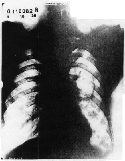
Radiograph II, taken at our Clinic on October 20, 1938 and showing extension of the tubercular process.

The prognosis on the first film dated September 16th, 1938, would indicate a very serious situation.
The prognosis on the third film dated June 18th, 1940, without knowing anything about the other two, would be very good.”
In the Spring of 1942, Mr. S. M. went to work for Fisher Body, a division of General Motors Corporation. He did hard manual labor, worked long hours and overtime as well. During his employment he was given examinations and X-Rays were taken. They established his good health and he was permitted to continue to work. His sputum has been negative since his recovery. He has continued working and testified in the Federal Court Trial in 1946.
Radiograph III, taken on July 8, 1939, about nine months after Treatment and showing healing of the cavity and the lung tissue undergoing reconstruction.

EXTENSIVE MILIARY
TUBERCULOSIS
With
Tubercular Meningitis and Tubercular Nephritis and Splenitis
CASE No. 48
The rapid advance of the disease in Miss N. A., age 14, when treated by the writer in July 1922, was reversed from the terminal stage by one injection. She had been in the Detroit Tuberculosis Sanitarium from the end of January until April. The radiographic findings of January 30, 1922, were:
“Both diaphragm leaves are clear. The trachea shows no compression or deviation. The heart shadow is normally placed. Increased deposit at the hilum on both sides. Some accentuation of the linear markings throughout the right lung field. No definite evidence of a parenchymal lesion in the right lung field. There is extensive parenchymal infiltration throughout the left lung field, with greatest changes in the upper half. Several small annular shadows at the apex and at the level of the first and second ribs anteriorly. We believe these represent small cavity formations. Diagnosis: Advanced parenchymal tuberculosis confined to the left lung field.”
It will be noted that the lesions are located subclavicularly as in the most rapidly disseminating type. Six months later I examined her. She was emaciated, bedfast, comatose, with frequent projectile vomiting for three weeks, cyanotic, with rapid shallow respiration, very rapid, thready, practically uncountable pulse, with the head drawn back in continuous opisthotonus. The fever was 105.5°F. The heart was drawn over into the right side of the chest; the left chest was empty of viscera so far as could be determined, but contained fluid that splashed on shaking her. This lung had spontaneously ruptured.
The right lung showed huge cavitations and consolidations, and the splenic area presented a hard, fixed tumefaction as large as her head. It could not be determined then if this was kidney, spleen or both involved in the tuberculosis process. She was also near death with a tubercular meningitis. Two micro-micrograms of the Synthetic Survival Reagent were given intramuscularly. The recovery process was slow at first, but this was to be expected considering the condition she was in at the time I examined her. It was some months before she got out of bed. A year later the heart was still on the right side of the chest, but the left chest was not empty anymore, it was full of apparently a fluid and fibrotic tissue. At the time she was walking about, her heart rate was still exceedingly rapid and weak, though it had dropped from around 150 to 130 per minute. Her temperature was normal. After that she gradually got better and the heart went gradually over into the left chest where it belonged. The heart rate slowed down.
An X-Ray taken July 24, 1944, shows that the left chest, which was so badly involved, is not exactly normal yet in all these twenty-two years because there is less lung tissue there. That is because part of the chest is replaced by the structures of the mediastinum. The lung tissue shows markings in chests that one would interpret as healed tuberculosis. These marks are very, very small, about a millimeter or two in diameter, dense, fibrous, showing calcification. It shows that the healing process has been very complete, and the scar tissue present from the large lesions healed and are very, very minute.
In this case we have a recovery with reconstruction in both lungs. The other pathological conditions cleared up during the recovery process. Her weight has increased to 145 pounds. She is married and has given birth to twins, pretty husky children. She has remained well to date (January, 1957).
The large mass in the splenic area disappeared completely, but the first improvement was noted in the nervous system, the disappearance of the opisthotonus and comatose state. This showed the SSR had taken effect, but still it was doubted that so much lung destruction could ever be repaired. Time showed that the advantage given here by the SSR not only permitted full lung reconstruction and return of the abdominal lesion to normal, but a general excellent health was gained that proved resistant to the usual infections. Her twins showed a remarkable immunity to the usual school children infections that none other exhibited, indicating a possible gene change.
FAR
ADVANCED TUBERCULAR PNEUMONIA
Or Acute
Military Invasion, in Extremus at Time of Treatment
CASE No.49
Another case of pulmonary tuberculosis in extremus at the time of Treatment with a dose of SSR is that of Mrs. M. B. and is of the type that has always advanced to fatality, namely originating in subclavicular lesions in both lungs. At the time of our Treatment April 2, 1934, her condition was too critical to permit a thorough differentiation as to whether she was dying of an acute tubercular pneumonia, or of an acute widespread military tuberculosis. She was “sunk in bed,” fever hovering about 104°F., rapid, thready pulse, cyanosis, with flush from the fever, yet the skin showed yellow hemolytic color after compressing the flushed capillaries. The breathing was very shallow and rapid, and very little but bloody sputum was raised. The physical findings scarcely revealed the cavity, only rales and solidification. No radiograph was taken at the time. A 2 cc. injection of the SSR, 10-(12), was given immediately. She had progressed to this stage from an early subclavicular and apical involvement thought to be a “cold” in August 1931, when she entered the Herman Kiefer Hospital of Detroit. The condition advanced to that shown in their film of April 23, 1932, Radiograph I. She remained in the two Detroit Public Tuberculosis Hospitals up to late in March 1934, when she was brought to our Clinic. Films of January 18, 1934, and March 8, 1934, show the advance of the disease under their care with the development of the large, shaggy cavity with slight fluid level behind the right clavicle. The latter film being reproduced here, Radiograph II. The experts offered to perform a Thoracoplasty operation upon her at that time. She left because she did not want this operation. She felt too sick for such a drastic operation, with her high fever and exhaustion. Her sister brought her to our Clinic on March 31, 1934. The sputum was loaded with tubercle bacilli. The films show the advance of the disease in both lungs and the infiltrative development of the cavity wall.
This report shows the condition of Mary B. when she entered the hospital.

Radiograph I, made in April 1932, shows advance of disease from August 1931, in the hospital.

The films show the advance of the disease in both lungs and the infiltrative development of the cavity wall. We took no radiographs until six months later when she was up and about and doing light work. Radiograph III, September 24, 1934, shows a smooth wall cavity. At this time there were no tubercle bacilli found in the sputum at daily tests for two weeks.

Radiograph No. II., shows advance of disease in hospital up to March 8, 1934, with cavitations.
This cavity represents the area where the infection had taken place, and where lung tissue was again healing in after the disease tissue was removed, and the completed process is shown in Radiograph IV, September 12, 1942.
She left the city after leaving our clinic and within a year went to work, at which she has remained ever since besides being married and living a normal life. Frequent sputum tests showed no more germs of tuberculosis and her gain in weight and perfect health mark her cure as complete. Physical findings are normal. Her X-Ray report at the time of entering the Herman Kiefer Hospital is reproduced in Photostat, from the Court Documentations. Reports in June 1960 show persisting recovery, good health, and working regularly. (See Appendix).

Radiograph No. III, shows the cleaning out of the tubercular infection and a sterile cavity undergoing healing. This radiograph was taken on September 24, 1934, about 5 ½ months after Treatment.
Radiograph IV, showing the cured state in 1942, eight years after Treatment.

ADVANCED
TUBERCULOSIS
With
Cavitations in Both Lungs
CASE No. 50
This is a case of bilateral cavitation with subclavicular lesions and two large thin walled apical cavitations on the right side. She steadily deteriorated under the best sanitarium care in Cleveland, Ohio (Sunny Acres), and in Detroit, Michigan, from 1931, when her thyroid gland was removed at the Crile Clinic of Cleveland, until September 2nd, 1934, when she received Treatment in our Clinic at Detroit.

Miss C. P. was examined at Herman Kiefer Hospital, Detroit, Michigan, in March 1932. A Photostat of the hospital X-Ray report is reproduced here. This is done because the radiograph does not show all of the lesions well.
Radiograph I shows the extent of the disease on February 24, 1934, about six months before she came to us for Treatment. Phrenectomy and pneumothorax had been unsuccessful in curing this patient. At the time we first saw her, the prognosis was serious. Bloody sputum loaded with tubercle bacilli was expelled up to the time of our examinations in September 1934. We found the upper half of both lungs invaded and a highly toxic state that resulted in unusual muscular weakness. On September 2, 1934, she was given 2 cc’s of the serially arranged SSR Carbonyl groups. This toxic state quickly left so that two weeks later she went to work instead of being confined to bed rest. She received a second Treatment on November 24, 1935. Since then she has had three more Treatments over the years, one in 1937, 1939, and 1942. She has been working, is married and in good health, according to our last report. Radiograph II, taken March 22, 1943, shows the cured state, with minimum of scar tissue and return of normal lung parenchyma.
This case shows how the basic toxic state that caused hyperthyroidism requiring thyroidectomy, also removed her resistance against tuberculosis infection, and caused other disturbances resulting in muscular weakness. This latter effect persisted until the time of receiving the Carbonyl Therapy, and then very quickly disappeared. It was no doubt due to suprarenal cortex inhibition. Her statement on her weakened condition includes this: “When I first visited Dr. Koch, I was very short of breath and was able to walk a short distance only,” and “I felt better almost immediately after the Treatment, and went back to work fourteen days later, September 16th.”
The testimony of Dr. Hague on this patient’s X-Rays has been paraphrased and reproduced here.
After examination of the X-Rays from Sunny Acres Sanitarium, Dr. Hague stated in regard to the X-Ray taken 8/6/32 that: “This is a radiograph of a thorax of a female patient, showing the chest cavities with a tuberculous process in the upper half of both lungs, of an advanced degree, with cavitations in the left apex, about three of them contiguous with one another, so I shall measure them all together. They measure two inches by one inch.
“There are also some shadows in the opposite side that suggest smaller cavitations behind the second rib anteriorly on the right side. This area of whiteness is an extensive tuberculosis process in the upper lobe of the right lung. A similar condition exists on the left side, but it doesn’t show so much density as on the right, because there is a cavitation process which has taken away some of the fibrosis and that has been spat out as sputum.”
“The descending bronchi are thickened because of repeated drainage from the upper areas of infection that have passed down into the lower trunks on both sides — lower bronchial trunks on both sides.”
X-Ray film dated 11/5/32: “This film shows the same patient with an aggravation of the disease, in which there is a shadow in the first interspace anteriorly, suggesting cavitation; and an enlargement of the shadows on the left side, indicating enlargement of the previous cavities. And, I believe an angular shadow that is on the left side in the previous film now shows more clearly that it is becoming a cavity, too. So that you now have an area of potential multiple cavitations measuring three inches by two inches. I would say that this patient is worse on this (11/5/32) film than on that one (8/6/32).
“X-Ray film dated 2/11/33: “The tuberculosis process in the right lung has increased. The cavitation is large; measures an inch and a quarter outside measurements in both diameters. The total area of cavitation in the left upper lung is slightly more, but there is a concurrent factor of a pneumothorax in which there is air in the base and up over the upper lobe of that lung.”
X-Ray film dated 5/17/33: “The only significant change in these two sets of film is that there is a little better compression over the apex of this lung, and one fairly strong adhesion band at a level of the third rib anteriorly is holding lung structure from complete collapse in that area.”
X-Ray film dated 8/16/33: “The same conditions exist in this film, with the exception that the outline of cavity in the right upper lobe, measuring two by one inches, is more clearly seen. There are still adhesions on the left side.”
X-Ray film dated 11/29/33: “I would say the left lung doesn’t show any significant change from the left lung in the immediately preceding, but the changes in the right upper lobe indicate the cavitation a little more sharply outlined, and a little heavier in cavitation wall thickening, which would suggest to me that there is more activity (the tuberculous process would be more active, creating more inflammation), and the response to the inflammation is characterized by a deposit of fibrous tissue surrounding that cavity.”
X-Ray film dated 2/24/34: “This is a little lighter film. The right apical cavity is clearly seen. The fibrous tissue surrounding it is a little less in density, but it is still present.”
X-Ray film dated 5/26/34 was then shown to Dr. Hague and he stated his opinion as to the general picture of the pathological condition of this patient at the time of this picture. “The three last views show no appreciable improvement under pneumothorax therapy. The diseased area of the left lung, with its cavitations, has not completely collapsed, because there are adhesions remaining, and the cavitation in the right apex with the associated fibrosis still exists, and I would say that that is a very serious case of active tuberculosis.”
Where the patient is still showing positive sputum, the prognosis is serious. (It should be noted that this last X-Ray (5/26/34) was taken over 3 months before the author treated the patient.)
Dr. Hague was shown an X-Ray taken about seven and one-half years after the patient was first treated in Dr. Koch’s Clinic. He stated that the X-Ray taken February 19, 1942 “indicates a very marked improvement of this patient. The pneumothorax previously seen has now disappeared, the gas has been absorbed, and the lung has re-expanded to fill the chest cavity. The areas of former large cavities in this side, in the left upper first and second interspaces, have practically gone, and the annular shadow on the right side in the first interspace also is gone, but there still remains a mild fibrosis in the first and second interspace at the site of the previous infection. There are fairly heavy hilar shadows in the left upper mediastinal area which have come from the inflammatory reaction of the large area of cavitation previously seen.”
The last film that had been taken, on March 22, 1943, is reproduced here and is Radiograph II. Dr. Hague stated: “That it does not show very much change from the one immediately preceding (2/19/42). I would say it is about stationary.”
To the question: “Assuming at the time of the last two films that the general health of the patient was good, and there was no sputum or no blood, what would you say as regards improvement or recovery process in these films?” Dr. Hague answered: “Well, having seen all the cavitations of the left side and the large one on the right — these two show a remarkable removal of disease process. It would be considered an excellent recovery if it were in the ordinary course of observation in a sanitarium. We would consider that a cure, under sanitarium conditions.”

Radiograph No. I, taken February 24, 1934, about six months before Treatment, showing cavitations in the right apex.

Radiograph No. II, taken March 22, 1943, showing recovery. It was taken for Court purposes.
TUBERCULOSIS OF
LUMBAR SPINE
CASE No. 51
J. A., age four, came to us with a diagnosis of tuberculosis of the spine in August 1924. He had been diagnosed by Detroit’s leading Orthopedist, Dr. La F----, Sr., who advised an Ablee Splint operation. He was supported by a brace that limited motion and reduced the pain only partially.
The family history showed his older sister had far advanced pulmonary tuberculosis.
For about a year he had increasing pain in the lower back and legs, found it difficult to get up after falling and would cry out with pain when he relaxed in bed on going to sleep. We fitted him with a brace, too. It did not limit his motion too much, but it did aid him during his sleep.
Examination showed a typical tuberculosis kyphosis in the lower lumbar spine. The two lower vertebrae showed a sharp angulation that protruded with collapse of the bodies of the vertebrae. There was considerable swelling of the soft tissues and muscle spasm about the region, which limited motion.
We gave him 1 micro-microgram of the SSR serial system of Carbonyl groups. There was a steady improvement so that he could discard the brace entirely in November 1925. The kyphosis gradually disappeared and the pain and limitation of motion disappeared with it. Within a year the spine was straight.
The radiographs made by the Orthopedist, at the time, showed the collapse of the bodies of the fourth and fifth lumbars and the angulated deformity. The radiographs taken after he was cured more than eighteen years showed the two vertebrae fused in perfect alignment so as to form one vertebra. In the upper thoracic spine an area or rarefication the size of a large pea was to be seen. This showed in the healed spine as an area of dense bone where the disease was cured before it could do any damage. The radiographs showing the healed state were submitted as exhibits to the Federal Trade Commission, but we did not receive them when the exhibits were returned and thus we are unable to reproduce them here. In April 1957, two radiographs were taken of the lower spine; one an anterior-posterior view and the other a lateral view. The perfect alignment in all directions is seen. The spine is found now to be one vertebra short.
In high school he participated in sports and received a letter in track. He attended the University of Michigan. Since his graduation, he has continued to live an active life and earn his own living. In April 1957, he reported that he is in excellent health and that his back never bothers him.
Normally, tuberculosis of the thoracic vertebrae usually results in severe deformity under conventional methods of treatment, i.e., by holding the patient in a rigid shell for many years. Where there is present a metastatic lesion as in this case, the chances of survival are only slight.

Radiograph I, was taken in April 1957, of Mr. J. A. and shows the anterior-posterior view of the lower spine.

