Sirayam
PANDEY, et al.
Dandelion & Long Pepper Fruit vs Cancer
Dandelion & Long Pepper Fruit vs Cancer
http://yournewswire.com/weed-destroys-cancer/
University Study: Common Garden Weed Destroys Cancer
Cells In 48 Hours
by Baxter Dmitry
by Baxter Dmitry
Back in 2009, a group of Canadian researchers from the University of Windsor in Ontario started investigating a common weed as a potential cure for cancer, and the results are more than amazing.
It all started when an oncologist came across something quite interesting with some cancer patients. Believe it or not, the plant we are talking about is the common dandelion!
A post-doctoral fellow at the University of Windsor named Pamela Ovadje has done an extensive work on the topic. She dealt with the anti-cancer properties of dandelion and similar extracts.
According to Ovadje, “We had information from an oncologist, a collaborator here in Windsor, who had patients that showed improvement after taking dandelion root tea. And so, with a phone call, we decided to start studying what was in this tea that made patients respond to it, so we started digging up dandelions.”
She was quite suspicious in the beginning, but not because it was an all-natural source. “I figured dandelions are everywhere, and if there was something to it, people would have found this out already, “ she explained.
We should be glad to hear that the researchers have started conducting studies on dandelion root extract and its effects on cancer, as the results are astonishing.
“Since the commencement of this project, we have been able to successfully assess the effect of a simple water extract of dandelion root in various human cancer cell types in the lab and we have observed its effectiveness against human T-cell leukemia, chronic myelomonocytic leukemia, and pancreatic and colon cancers, with no toxicity to non-cancer cells. Furthermore, these efficacy studies have been confirmed in animal models (mice) that have been transplanted with human colon cancer cells.” [ http://www.uwindsor.ca/dandelionrootproject/ ]
Dandelion root extract was approved for human trials in February 2015. Now, it is in Phase 1 trials for end-stage blood-related cancers, such as leukemia and lymphoma.
According to Dr. Siyaram Pandey, professor of chemistry and biochemistry at the University of Windsor and principal research investigator for the project, dandelion root extract has quite a “good potential” to cause a death of cancer cells.
How Does it Work?
This extract causes cancer cells to go through apoptosis, a natural cell process where a cell activates an intracellular death program because it isn`t needed anymore. In brief, dandelion root extract causes the cancer cell to “commit suicide” without affecting the healthy ones.
Two cells perform apoptosis which is far better than chemotherapy drugs which kill one healthy cell for every 5 to 10 cancer cell, the dandelion extract.
It is important to mention that the concentration of this extract is much higher than the one which is currently available. Even though trials are still underway, this extract may be the future of cancer treatment!
The Dandelion Root Project is aimed at showcasing scientific evidence for the safe and effective use of dandelion root extract and other natural health products for cancer therapy.
The Dandelion Root Project started in 2009 in a bid to investigate the anticancer effect of the root extract of dandelions against cancer cells in the lab (in cells and in animal models). This project started with funding from the Knights of Columbus, Chapter 9671 (Windsor) and has been sustained by funding from other sources, including Seeds4Hope Grant (local Cancer Foundation), Lotte & John Hecht Foundation, The Pajama Angels and the Jesse & Julie Rasch Foundation. Private and personal donations have been made from the Windsor local community, as well as from all around Canada. We dedicate this project in the memory of Mr. Kevin Couvillon, who lost his battle with leukemia in 2010.
Since the commencement of this project, we have been able to successfully assess the effect of a simple water extract of dandelion root in various human cancer cell types, in the lab and we have observed its effectiveness against human T cell leukemia, chronic myelomonocytic leukemia, pancreatic and colon cancers, with no toxicity to non-cancer cells. Furthermore, these efficacy studies have been confirmed in animal models (mice) that have been transplanted with human colon cancer cells.
We also applied for Phase I clinical trials in 2012 for the use of DRE in hematological cancers and in November 2012, we obtained approval for the administration of DRE in human patients and currently, the dandelion root extract is under Phase 1 clinical trials for drug refractory blood cancers.
Studies to understand how dandelion root extract can identify differences between cancer cells and non-cancer cells are underway, while at the same time, the identification of the active components within the extract is ongoing. We are excitingly awaiting the results from these studies.
http://windsorstar.com/news/dandelion-root-cancer-treatment-enters-clinical-trials-company-to-market-it-formed
17 February 205
Dandelion
root cancer treatment enters clinical trials, company to
market it formed
Chris Thompson
Chris Thompson
To mark what would have been Kevin Couvillon’s 31st birthday, the revolutionary dandelion root natural cancer treatment program that bears his name had a few surprises to reveal Tuesday.
After overcoming some hurdles, Phase 1 of the clinical trials approved by Health Canada in 2012 have begun to proceed at the Windsor Regional Cancer Centre and 30 patients from across Canada are being sought.
Also, a new company to be known as Windsor Botanical Therapeutics Inc. has been formed to commercially market the product.
“This is one way for us to dedicate all of our work to Kevin Couvillon,” said Dr. Siyaram Pandey, the University of Windsor biochemist who oversees the Kevin Couvillon Research Project on the Anti-Cancer Effects of Dandelion Root Extract.
Friends and family of Couvillon, University of Windsor researchers and interested members of the medical community from as far away as Toronto gathered at the Dr. Murray O’Neil Medical Education Centre to mark Couvillon’s birthday, which was Sunday.
“He showed us nothing but strength through his 3½-year battle,” said an emotional Donna Couvillon, Kevin’s mother.
“Through all this and more, he believed he would be saved. Tragically, it was not to be.”
Couvillon, a talented sound engineer and musician, died on Nov. 24, 2010, after battling leukemia and the secondary infections caused by the treatment.
In 2011 Couvillon’s parents donated $20,000 to the dandelion root research, and on Tuesday a new scholarship in Kevin’s name was also announced.
Recent doctorate graduate Pamela Ovadje, one of the lead researchers in the project, outlined recent progress, including having reports published in a number of science journals.
She also said the project recently partnered with Calgary company Advanced Orthomolecular Research for quality-controlled production of the dandelion root extract.
“We’ve got the first batch ready for clinical trials,” said Ovadje.
The clinical trial will be focused on sufferers of blood-borne cancers such as leukemia who have tried all other avenues of treatment.
“This is for people who have exhausted all other options,” said Pandey.
Patients can be from across Canada, but treatment will be provided out of the Windsor Regional Cancer Centre through oncologist Dr. Caroline Hamm.
Hamm discovered the interesting properties of dandelion root while treating an 85-year-old woman with leukemia who saw a dramatic drop in her white blood count after drinking dandelion tea.
Tests in petri dishes and in mice have shown the dandelion extract attacks the cancerous cells, but does not impact healthy ones. This could provide an effective alternative to traditional treatments such as chemotherapy and radiation which cause collateral damage to the patient.
Details of the trial can be found at canadiancancertrials.ca and searching “dandelion.”
Also at Thursday’s event was the announcement of the creation of Windsor Botanical Theraputics Inc. to market the dandelion root extract.
“The main purpose of any company such as this is to bring the research and technologies to the market,” said CEO Dr. Joseph Elliott.
“We are the next step in bringing Dr. Pandey’s research to where patients can actually benefit.”
There are many dandelion natural health products on the market currently which can’t claim the anti-cancer benefit. Should the clinical trials reflect a benefit, the company would then be able to market on that basis, Elliott said.
“We will begin with standard claims, then cancer claims,” Elliott said.
The product will be available in capsule form.
http://canadiancancertrials.ca/trial/Default.aspx?dsEndecaNav=Ntk%3AMain_en|dandelion|1|%2CRo%3A0%2CNrc%3Aid-30-dynrank-disabled|id-130-dynrank-disabled|id-131-dynrank-disabled|id-132-dynrank-disabled|id-619-dynrank-disabled|id-620-dynrank-disabled|id-621-dynrank-disabled|id-622-dynrank-disabled&TrialId=OCT1226&lang=en
Dandelion
Root Extract in Patients with Refractory Hematologic
Malignancies
Summary:
Thirty patients with refractory (end stage) blood related cancers (lymphoma, leukemia, myelodysplastic syndromes) will be entered into the protocol in groups of three. Each subsequent group of three will have an increase in the dose of DRE until unacceptable side effects are experience by at least two out of the three in that group. Unacceptable side effects include admission to hospital for side effects related to the drug, death or inability to consume the recommended dose. Once dose limiting toxicity is identified, we move back to dose level previous to the one that causes the serious event, and that will be the recommended dose.
Trial Description
Primary Outcomes:
To define the recommended dose of DRE consumed orally.
Secondary Outcomes:
To evaluate the toxicity of DRE and their reversibility, association with dose in this same patient population.
To assess preliminary evidence of antitumour effects in those patients with measureable disease by documentation of objective responses using NCI criteria in patients with refractory hematologic malignancies who are receiving single agent DRE.
To measure the effect of the DRE on caspase activity in the blood cells of leukemia patients receiving the DRE. Correlation to know molecular factors will be performed where available (eg JAK2, cytogenetics, PLK-1).
Variables to be assessed include concomitant medications, concomitant complementary medicines, patient co-morbidities, number of prior therapies and the type and stage of cancer.
The design of this dose-finding phase I clinical trial was chosen to assess the DRE in the treatment of patients with refractory hematologic malignancies. Patients will be eligible for this study if they have a refractory hematologic malignancy and have exhausted all standard medical treatments for their cancer. Patients must not be pregnancy and must be at least 18 years of age and not over 70 years of age. Patients cannot have significant comorbidites that would increase the risk of death or toxicity from this product. They must be capable of adhering to the protocol.
The starting dose of DRE will be 1000 mg daily which will be provided by Dr. Pandey's lab at University of Windsor. Patient will receive their supply of DRE at the beginning of each week. This product will be dissolved in 125 cc of cold water or hot water and consume this in less than 1 hour. Honey, sugar or sweetener may be added to the product to improve tolerability. There is no premedication. The first 3 patients will start off at level 1 with 2 aliquots per day, taking 1 in the morning and the 2nd in the evening. The rate of subject entry and escalation to the next dose level will depend on assessment of the safety profile of patients entered at the previous dose level. Toxicity will be evaluated according to NCIC CTCAE v 3.0. Three patients within a dose level must be observed for one cycle (28 days) before accrual to the next higher dose level. Patients will remain on the DRE until progression of disease or until they develop unacceptable toxicity or withdraw from the study if they choose.
US2015010665
Medicament Containing Taraxacum Plant Root Extract for Treatment or Prevention of Cancer, and Method for Preparing Same
Medicament Containing Taraxacum Plant Root Extract for Treatment or Prevention of Cancer, and Method for Preparing Same
Inventor: Sirayam PANDEY, et al
The present invention relates to an improved method for preparing a medicament comprising a Taraxacum plant root extract for the treatment or prevention of a cancer. In one aspect, the method comprises freezing Taraxacum plant root to obtain a frozen root stock, said freezing step being selected to effect at least partial disruption of one or more root cells; dry grinding the frozen root stock to obtain a ground root powder, wherein during said dry grinding step the frozen root stock is maintained at a grinding temperature below about 40 DEG C.; steeping the ground root powder with a solvent to obtain a suspension having a liquid extract portion and a solid particle portion; and separating the liquid extract portion from the solid particle portion to provide a separated liquid extract for use in the medicament.
RELATED APPLICATIONS
[0001] This application claims the benefit of 35 U.S.C. 119(e) to U.S. Provisional Patent Application Ser. No. 61/597,453 filed on 10 Feb. 2012.
SCOPE OF THE INVENTION
[0002] The present invention relates to an improved method of preparing a medicament which includes a root extract of plants belonging to the genus Taraxacum, and which is for treatment, amelioration or prevention of cancers. More particularly, the present invention relates to the preparation of a pharmaceutical composition which includes Taraxacum plant root extracts for use in the treatment and/or prevention of cancers, and preferable colon cancers, pancreatic cancer, skin cancers such as melanoma, and blood cancers such as chronic lymphoid leukemia, chronic myeloid leukemia, chronic monocytic myeloid leukemia and Hodgkin's lymphoma.
BACKGROUND OF THE INVENTION
[0003] Plants of the genus Taraxacum, also commonly known as dandelions, are members of the Asteraceae family. These plants are commonly found in temperate zones of the Northern Hemisphere, and species of dandelions include T. officinale, T. erythrospermum, T. albidum, T. japonicum, T. laevigatum, T. erythrospermum and T. californicum.
[0004] Dandelions are tap-rooted biennial or perennial herbaceous plants with an average length of 15 to 30 cm. The leaves are large, light to dark green in color and cluster in a rosette at the base of the plant. The flowering stalks are upstanding and carries a solitary, terminal inflorescence. The florescence ranges from 7 to 15 mm in diameter and is composed of 140 to 400 yellow, ligulate florets. The fruits are conical achenes, brown and crowned by a white, hairy papus, which allows the seeds to be distributed by wind over long distances.
[0005] Taraxacum plant roots often contain a variety of compounds including sesquiterpenes, carotenoids, coumarins, flavonoids, phenolic acids, polysaccharides, eudesmanolides, triterpenes, sterols, steroids and others. Specific examples of such compounds include germacranolide, eudesmanolide, guaianolide, taraxacin, phenylpropanoid glycosides, taraxacoside, lactupircin, lutein, violaxanthin, esculin, scopoletin, quercetin, luteolin, rutin, chrysoeriol, caffeic acid, vanillic acid, syringic acid, ferulic acid, chlorogenic acid, chicoric acid, p-hydroxyphenylacetic acids, p-hydroxylbenzoic acid, inulin, glucans, mannans, prunasin, 11β, 13-dihydrolactucin, ixerin D, ainslioside taraxinic acid, β-glucopyranosyl, taraxinic acid, glucosyl ester, 11, 13-dihydrotaraxinic acid, 1′-glucoside, lactucopicrin, lactucin, cichorin, tetrahydroridentin B, taraxacolide-O-β-glucopyranoside, prunasin, dihydroconiferin, syringin, dihydrosyringin, taraxasterol, ψ-taraxasterol, homo-taraxasterol, stigmatsterol, cycloartenol, umbelliferone, taraxalisin, α-amyrin, β-amyrin, arnidiol, faradiol, lupeol, taraxol, taraxaserol, 3β-hydroxylup-18-ene-21-one, β-sitosterol, campesterol, lettucenin A, choline, mucilage, pectin, and taraxerol.
[0006] Dandelion extracts have been used in the past as for example antioxidants, diuretics, analgesics, anti-coagulants and anticancer agents. The publication “Evaluation of aqueous extracts of Taraxacum officinale on growth and invasion of breast and prostate cancer cells” International Journal of Oncology 32 (2008): 1085-1090 to Sigstedt reports on the anticancer activity of crude extracts prepared from the leaves (“DLE”), flowers (“DFE”) or roots (“DRE”) of the dandelion species Taraxacum officinale. The crude dandelion extracts in Sigstedt were prepared by 1) soaking 75 g of dried plant parts in water for 24 hours at room temperature; 2) filtering the resulting mixture to remove particulate matter; and 3) lyophilizing the mixture to obtain a powder. Sigstedt observes that DLE reduced the growth of MCF-7/AZ breast cancer cells, and not that of LNCaP C4-2B prostate cancer cells; and that both DFE and DRE failed to influence cancer cell proliferation.
[0007] The publication “Anti-carcinogenic Activity of Taraxacum Plant. I” Biol. Pharm. Bull. 22.6 (1999): 602-605 to Takasaki relates to dandelion root extracts prepared from the species Taraxacum japonicum. Takasaki describes extracting dried roots (600 g) of T. japonicum plant three times with 3 L of methanol for five hours each, and then evaporating the methanol solution to afford 109 g of a methanol extract. Takasaki additionally describes the preparation of a water extract obtained from extracting 60 g of T. japonicum roots with 0.38 L of water for 1 hour, and then lyophilizing the resulting solution. Takasaki describes that the methanol and water extracts inhibited initiation and promotion of two-stage chemical carcinogenesis.
[0008] In the separate publication “Anti-carcinogenic Activity of Taraxacum Plant. II” Biol. Pharm. Bull. 22.6 (1999): 606-610, Takasaki describes another dandelion root preparation of T. japonicum obtained from extracting dried roots (6.7 kg) with 40 L of n-hexane three times for 8 hours each to produce a 120.5 g extract.
[0009] Dandelion plant parts have been utilized to prepare extracts in various forms including capsules and tinctures. Dandelion roots in particular have been harvested for preparing “dandelion coffee” obtained by steeping dried ground plant root in boiling water. It has been appreciated that such conventional forms of dandelion extracts are ordinarily associated with lower anticancer activities, inducing as low as 10% cell death when introduced to a cancerous or tumor tissue.
SUMMARY OF THE INVENTION
[0010] It is an object of the present invention to provide a medicament or pharmaceutical composition for the treatment, amelioration and/or prevention of cancers, and which includes a Taraxacum plant root extract, preferably in combination with a pharmaceutically acceptable carrier, a diluent, a binding agent, an adjuvant and/or other anticancer agents.
[0011] A further object of the present invention is to provide a Taraxacum plant root extract which is suitable and/or beneficial for use as a medicament or human consumption.
[0012] A yet further object of the present invention is to provide a method of preparing a medicament or pharmaceutical composition having a Taraxacum plant root extract which includes one or more compounds useful for the treatment, amelioration and/or prevention of a cancer.
[0013] It has been appreciated that a Taraxacum plant root extract may be useful in the treatment and/or prevention of cancers, and which may include without restriction pancreatic cancers, colon cancers, blood cancers and skin cancers. Such skin cancers may be melanoma, and such blood cancers may be leukemia, such as but not limited to Hodgkin's lymphoma, chronic lymphoid leukemia, chronic myeloid leukemia and chronic monocytic myeloid leukemia.
[0014] In one possible method, a medicament for the treatment or prevention of a cancer may be prepared by: freezing Taraxacum plant root to obtain a frozen root stock, said freezing step being selected to effect at least partial disruption of one or more root cells; dry grinding the frozen root stock to obtain a ground root powder, wherein during said dry grinding step the frozen root stock is maintained at a grinding temperature below about 40° C.; steeping the ground root powder with a solvent to obtain a suspension having a liquid extract portion and a solid particle portion; and separating the liquid extract portion from the solid particle portion to provide a separated liquid extract for use in the medicament.
[0015] Although not intended to be bound by theory, it was experimentally shown that anticancer compounds contained in a Taraxacum plant root may undergo a reduction in their activities if subject to an elevated temperature in a dry environment, although such effect is less pronounced or absent in wet environments. The applicant has recognized that during an extract preparation process, a Taraxacum plant root may unfavorably be left exposed to dry heat, resulting in the reduction and possibly elimination of anticancer activities. A Taraxacum plant root and its anticancer activity may be most vulnerable to the deactivating effects of dry heat during the grinding step where the plant roots, root cells and cellular contents could be heated on contact with a rotating element of a grinder, such as a grinder blade. A number of experiments were performed to show that loss of activities could occur above 40° C., and a complete loss of anticancer activities may result from exposure to a temperature above 70° C.
[0016] In a preferred embodiment, the grinding temperature is kept below about 0° C., more preferably below about −25° C., and most preferably below about 40° C.
[0017] Furthermore, it has been appreciated that the anticancer activity of medicament having a Taraxacum plant root extract may be improved if prepared with ground plant root obtained from dry grinding rather than wet grinding. Dry grinding is believed to provide improved and/or more controllable disruption of root cells, and thus greater amounts of intracellular anticancer contents or compounds available for subsequent extraction steps. Preferably, the frozen root stock is dry ground to an average particle size of less than about 100 μm, more preferably less than about 50 μm and most preferably between about 1 μm and about 30 μm.
[0018] In a preferred aspect, a Taraxacum root extract may be especially useful for inclusion in a medicament for the treatment, amelioration or prevention of cancers when prepared with dandelion roots obtained from dormant Taraxacum plants harvested before, or more preferably within about 90 days, and most preferably about 30 days prior to plant blooming or budding in the spring season, or before entering dormancy in the winter season when bud growth ceases.
[0019] Although not intended to be bound by theory, it is believed that Taraxacum plant roots undergo physiological changes in preparation of blooming or dormancy. Specifically, based on the experimental results obtained from the extracts prepared from dandelion roots (“DRE”) obtained at three different time points (Spring, Summer and beginning of Fall), the extracts prepared from the roots harvested in early spring and beginning of the fall period were shown to be the most effective in inducing cell death in cancer cells. In particular, dandelion roots harvested in the province of Ontario, Canada in March, September and October were shown to be highly effective in inducing apoptosis of cancer cells. It is believed that the anticancer compounds in the root extract are synthesized in preparation for dormancy (during the cold weather), and which may be involved in inducing cell death and eliminating the aged cells in the plant in preparation for winter.
[0020] In a preferred embodiment, the Taraxacum plant root is, prior to the freezing step, dried to a relative humidity of about 5% to 10%. Preferably the plant root is diced into root pieces, which may have an average dimension between about 0.2 cm and 1.0 cm.
[0021] The Taraxacum plant root is preferably obtained from a Taraxacum species including but not limited to T. officinale, T. erythrospermum, T. albidum, T. japonicum, T. laevigatum, T. erythrospermum and T. californicum. Most preferably, the plant root is harvested from T. officinale or T. laevigatum collected from an open grassy area.
[0022] Preferably in the freezing step the plant root is contacted or submerged in liquid nitrogen, or alternatively, subjected to a freezing temperature below 0° C., or more preferably between about −210° C. and about −30° C., for about 5 minute to 24 hours or until substantially frozen.
[0023] The dry grinding step may be carried out with a grinder, including but not limited to a mortar and pestle, a pulverizer, an impingement grinder and a micronized milling machine to effect substantial disruption of root cells. To reduce exposure to elevated temperatures in a dry environment above 40° C., the grinder is preferably cooled, with for example liquid nitrogen, to prevent heating on contact with the frozen root stock or the resulting ground root powder. Preferably, the grinder is cooled below about −25° C., and more preferably below about −50° C.
[0024] To better effect the release of therapeutically active compounds located inside the root cells, the grinding step is most preferably performed to disrupt or break open the cells and release their inner contents.
[0025] The ground root powder is steeped or soaked in a liquid or solvent, preferably in a polar solvent, such as water at a soaking temperature between about 5° C. and about 100° C., or most preferably at about 25° C. Other suitable solvents include but not limited to pentane, cyclopentane, cyclohexane, benzene, toluene, 1,4-dioxane, chloroform, diethyl ether, dichloromethane, tetrahydrofuran, ethyl acetate, acetone, dimethylformamide, acetonitrile, dimethyl sulfoxide, propylene carbonate, formic acid, n-butanol, isopropanol, n-propanol, ethanol, methanol and acetic acid. The ground root powder is preferably soaked in the liquid between about 5 minutes to about 24 hours, or more preferably between about 10 minutes and about 30 minutes with or without stirring. Most preferably, the ground root powder is soaked in water at 10 g ground root 50 mL water, and is boiled between 10 minutes and 30 minutes.
[0026] The liquid extract portion of the suspension may be separated from the solid particle portion preferably by filtration and/or centrifugation. Preferably, centrifugation, if performed, is carried out between 5000×g to 8000×g to remove any excess fibers. Filtration is preferably performed using suction filtration and a paper filter. The paper filter preferably has the pore size of less than or equal to about 0.45 μm, and most preferably less than or equal to about 0.22 μm. In a most preferred embodiment, the filtration step is performed stepwise using paper filters of decreasing pore sizes (such as 0.45 μm filter, followed by 0.22 μm filter). One or more filters or filter papers utilized for the filtration step may be configured to remove a bacteria.
[0027] The separated liquid extract obtained from the suspension is preferably freeze dried to an extract powder. Preferably, the freeze drying step is performed at a temperature between about −80° C. and −40° C.
[0028] The extract powder may be included in the medicament together with a pharmaceutically acceptable carrier, a diluent, a binding agent, an adjuvant and/or additionally anticancer agents. Such anticancer agents may include metformin, hydroxyurea, cyclophosphamide and/or etoposide.
[0029] The medicament preferably include a dosage form which contains the extract powder in a range about 5 mg/kg weight/day to about 1000 mg/kg weight/day, and preferably about 10 mg/kg weight/day to about 70 mg/kg weight/day. Alternatively, 0.2 to 200 g, preferably about 0.5 g to about 70 g, and most preferably about 1 to 4 g of the extract powder is preferably included in medicament form as a daily dosage.
[0030] In yet another aspect, the present invention provides a method for preparing a medicament comprising a Taraxacum plant root extract for treatment or prevention of a cancer, the method comprising the steps of: (1) freezing Taraxacum plant root to obtain a frozen plant root stock, said freezing step selected to effect at least partial disruption of one or more root cells, wherein said Taraxacum plant root comprises a dormant Taraxacum plant root harvested either prior to plant budding or blooming, or after cessation of bud growth; (2) dry grinding said frozen plant root stock to obtain a ground plant root powder with an average particle size of less than about 100 μm, and preferably less than about 50 μm, wherein during said dry grinding step the frozen root stock is maintained at a grinding temperature below about 40° C.; (3) soaking the ground plant root powder in a solvent comprising one or both of ethanol and water to produce a mixture having a liquid solution portion and a solid portion; (4) separating the liquid solution portion from the solid portion; and (5) freeze drying the liquid solution portion to obtain the Taraxacum plant root extract as a dried extract powder, and optionally mixing the dried extract portion with one or more of a pharmaceutically acceptable carrier, a diluent, a binding agent, an adjuvant and an anticancer agent.
[0031] The dormant plant root is harvested within 90 days, and preferably about 30 days, prior to first seasonal plant blooming or budding. The plant root may be from a plant belonging to a species of T. officinale, T. erythrospermum, T. albidum, T. japonicum, T. laevigatum, T. erythrospermum and T. californicum.
[0032] The plant root is preferably dried to a relative humidity of less than about 10% before freezing. In the following freezing step, the plant root is preferably contacted or submerged in liquid nitrogen to an average freezing temperature between about −210° C. and about −30° C.
[0033] In the dry grinding step, the frozen root stock is ground preferably to an average particle size of less than about 50 μm, and more preferably between about 1 μm to about 30 μm. The dry grinding step may be carried out with a grinder, including but not limited to a mortar and pestle, a pulverizer, an impingement grinder and a micronized milling machine to effect substantial disruption of one or more root cells. To reduce exposure to elevated temperatures in a dry environment, the grinder is preferably cooled, with for example liquid nitrogen, to a temperature below about −25° C., and preferably below about −50° C.
[0034] The solvent for use in the soaking step may additionally include one or more of pentane, cyclopentane, cyclohexane, benzene, toluene, 1,4-dioxane, chloroform, diethyl ether, dichloromethane, tetrahydrofuran, ethyl acetate, acetone, dimethylformamide, acetonitrile, dimethyl sulfoxide, propylene carbonate, formic acid, n-butanol, isopropanol, n-propanol, methanol or acetic acid. The soaking step is most preferably performed at a soaking temperature between about 5° C. and about 100° C., preferably for a period of about 5 minutes to about 24 hours, with or without stirring.
[0035] Various techniques may be utilized for separating the liquid solution portion from the solid portion in the mixture. Such techniques may include but not limited to centrifugation and filtration. Centrifugation, if performed, is preferably carried out between 5000×g to 8000×g. Filtration, if used, is most preferably performed at least twice using at least two filters of different pore sizes, such as about 0.45 μm and about 0.22 μm. For improved safety for human consumption, one or more filters or filter papers utilized for the filtration step may be configured to remove a bacteria.
[0036] In a preferred embodiment, the grinding temperature is below about 0° C., more preferably below about −25° C., and most preferably below about −40° C.
[0037] The medicament preferably include a dosage form which contains the Taraxacum plant root extract in a range about 5 mg/kg weight/day to about 1000 mg/kg weight/day, and preferably about 10 mg/kg weight/day to about 70 mg/kg weight/day. Alternatively, about 0.5 g to about 70 g, and preferably about 1 to 4 g of the Taraxacum plant root extract is preferably included in the medicament in a daily dosage form.
[0038] In aspect (1), the present invention provides a method for preparing a medicament for the treatment or prevention of a cancer, the method comprising: freezing Taraxacum plant root to obtain a frozen root stock, said freezing step being selected to effect at least partial disruption of one or more root cells; dry grinding the frozen root stock to obtain a ground root powder, wherein during said dry grinding step the frozen root stock is maintained at a grinding temperature below about 40° C.; steeping the ground root powder with a solvent to obtain a suspension having a liquid extract portion and a solid particle portion; and separating the liquid extract portion from the solid particle portion to provide a separated liquid extract for use in the medicament.
[0039] In aspect (2), the current invention provides a method according to aspect (1), wherein said cancer is a colon cancer, a pancreatic cancer, a blood cancer or a skin cancer.
[0040] In aspect (3), the present invention provides a method according to aspect (1) and/or (2), wherein said cancer comprises said blood cancer or said skin cancer, and is selected from the group consisting of chronic lymphoid leukemia, chronic myeloid leukemia, chronic monocytic myeloid leukemia, Hodgkin's lymphoma, and melanoma.
[0041] In aspect (4), the present invention provides a method according to any one or more of aspects (1) to (3) in any combination, wherein prior to said freezing step, the method further comprises drying said plant root to a relative humidity between about 5% to about 10%.
[0042] In aspect (5), the present invention provides a method according to any one or more of aspects (1) to (4) in any combination, wherein said Taraxacum plant root comprises a dormant Taraxacum plant root harvested either prior to plant blooming or budding, or after cessation of bud growth.
[0043] In aspect (6), the present invention provides a method according to any one or more of aspects (1) to (5) in any combination, wherein said dormant Taraxacum plant root is harvested within about 90 days, and preferably about 30 days, prior to said plant blooming or budding.
[0044] In aspect (7), the present invention provides a method according to any one or more of aspects (1) to (6) in any combination, wherein said Taraxacum plant root is from a plant belong to a species selected from the group consisting of T. officinale, T. erythrospermum, T. albidum, T. japonicum, T. laevigatum, T. erythrospermum and T. californicum.
[0045] In aspect (8), the present invention provides a method according to any one or more of aspects (1) to (7) in any combination, wherein said freezing step comprises contacting or submerging the plant root in liquid nitrogen, or freezing the plant root to an average freezing temperature between about −210° C. and about −30° C.
[0046] In aspect (9), the present invention provides a method according to any one or more of aspects (1) to (8) in any combination, wherein said dry grinding step comprises dry grinding the frozen root stock to an average particle size of less than about 100 μm, and preferably less than about 50 μm.
[0047] In aspect (10), the present invention provides a method according to any one or more of aspects (1) to (9) in any combination, wherein said dry grinding step comprises dry grinding the frozen root stock with a grinder selected from the group consisting of a pulverizer, an impingement grinder and a micronized milling machine, and wherein the grinder or a component thereof is cooled below about −25° C., and preferably below about −50° C., to prevent heating on contact with the frozen root stock or the ground root powder.
[0048] In aspect (11), the present invention provides a method according to any one or more of aspects (1) to (10) in any combination, wherein said solvent comprises one or more of water, pentane, cyclopentane, cyclohexane, benzene, toluene, 1,4-dioxane, chloroform, diethyl ether, dichloromethane, tetrahydrofuran, ethyl acetate, acetone, dimethylformamide, acetonitrile, dimethyl sulfoxide, propylene carbonate, formic acid, n-butanol, isopropanol, n-propanol, ethanol, methanol and acetic acid.
[0049] In aspect (12), the present invention provides a method according to any one or more of aspects (1) to (11) in any combination, wherein said steeping step comprises soaking the ground root powder in water at a soaking temperature between about 2° C. and about 150° C., and preferably 5° C. and 100° C., preferably for a period of about 5 minutes to about 24 hours, with or without stirring.
[0050] In aspect (13), the present invention provides a method according to any one or more of aspects (1) to (12) in any combination, wherein said separation step comprises at least one of filtration and centrifugation, and wherein the filtration is performed once or more than once using a plurality of filters of same or different pore sizes, and the centrifugation is performed at 5000×g to 8000×g.
[0051] In aspect (14), the present invention provides a method according to any one or more of aspects (1) to (13) in any combination, wherein said separation step comprises filtering the suspension at least twice with a first filter having a first pore size of about 0.45 μm and a second filter having a second pore size of about 0.22 μm, and wherein the second filter is selected to remove a bacteria.
[0052] In aspect (15), the present invention provides a method according to any one or more of aspects (1) to (14) in any combination, wherein prior to the dry grinding step, the method further comprises dicing said plant root to produce a plurality of root pieces.
[0053] In aspect (16), the present invention provides a method according to any one or more of aspects (1) to (15) in any combination, wherein said grinding temperature is below about 0° C.
[0054] In aspect (17), the present invention provides a method according to any one or more of aspects (1) to (16) in any combination, wherein said grinding temperature is below about −25° C., and preferably below about −40° C.
[0055] In aspect (18), the present invention provides a method according to any one or more of aspects (1) to (17) in any combination, wherein said method further comprises freeze drying the separated liquid extract to obtain an extract powder, and optionally mixing the extract powder with one or more of a pharmaceutically acceptable carrier, a diluent, a binding agent, an adjuvant and an anticancer agent.
[0056] In aspect (19), the present invention provides a method according to any one or more of aspects (1) to (18) in any combination, wherein said anticancer agent is one or more of metformin, hydroxyurea, cyclophosphamide and etoposide.
[0057] In aspect (20), the present invention provides a method according to any one or more of aspects (1) to (19) in any combination, wherein said medicament comprises a dosage form having the extract powder in a range of about 5 mg/kg weight/day to about 1000 mg/kg weight/day, and preferably about 10 mg/kg weight/day to about 70 mg/kg weight/day.
[0058] In aspect (21), the present invention provides a method according to any one or more of aspects (1) to (20) in any combination, wherein said medicament comprises a daily dosage form having the extract powder in a range of about 0.5 g to about 70 g, and preferably about 1 to 4 g.
[0059] In aspect (22), the present invention provides a method for preparing a medicament comprising a Taraxacum plant root extract for treatment or prevention of a cancer, the method comprising the steps of: (1) freezing Taraxacum plant root to obtain a frozen plant root stock, said freezing step selected to effect at least partial disruption of one or more root cells, wherein said Taraxacum plant root comprises a dormant Taraxacum plant root harvested either prior to plant budding or blooming, or after cessation of bud growth; (2) dry grinding said frozen plant root stock to obtain a ground plant root powder with an average particle size of less than about 100 μm, and preferably less than about 50 μm, wherein during said dry grinding step the frozen root stock is maintained at a grinding temperature below about 40° C.; (3) soaking the ground plant root powder in a solvent comprising one or both of ethanol and water to produce a mixture having a liquid solution portion and a solid portion; (4) separating the liquid solution portion from the solid portion; and (5) freeze drying the liquid solution portion to obtain the Taraxacum plant root extract as a dried extract powder, and optionally mixing the dried extract portion with one or more of a pharmaceutically acceptable carrier, a diluent, a binding agent, an adjuvant and an anticancer agent.
[0060] In aspect (23), the present invention provides a method according to aspect (22), wherein said cancer is a colon cancer, a pancreatic cancer, a blood cancer or a skin cancer.
[0061] In aspect (24), the present invention provides a method according to aspect (22) and/or (23), wherein said cancer is chronic lymphoid leukemia, chronic myeloid leukemia, chronic monocytic myeloid leukemia, Hodgkin's lymphoma, or melanoma.
[0062] In aspect (25), the present invention provides a method according to any one or more of aspects (22) to (24) in any combination, wherein said dormant Taraxacum plant root is harvested within about 90 days, and preferably about 30 days, prior to first seasonal plant blooming or budding.
[0063] In aspect (26), the present invention provides a method according to any one or more of aspects (22) to (25) in any combination, wherein said Taraxacum plant root is from a plant belonging to a species selected from the group consisting of T. officinale, T. erythrospermum, T. albidum, T. japonicum, T. laevigatum, T. erythrospermum and T. californicum.
[0064] In aspect (27), the present invention provides a method according to any one or more of aspects (22) to (26) in any combination, wherein prior to said freezing step, the method further comprises drying said plant root to a relative humidity less than about 10%.
[0065] In aspect (28), the present invention provides a method according to any one or more of aspects (22) to (27) in any combination, wherein said freezing step comprises contacting or submerging the plant root in liquid nitrogen to an average freezing temperature between about −210° C. and about −30° C.
[0066] In aspect (29), the present invention provides a method according to any one or more of aspects (22) to (28) in any combination, wherein said dry grinding step comprises dry grinding the frozen root stock with a grinder to the average particle size of less than about 50 μm, and to effect substantial disruption of said one or more root cells, the grinder being selected from the group consisting of a pulverizer, an impingement grinder and a micronized milling machine, and wherein said dry grinding step further comprises cooling said grinder to a temperature below about −25° C.
[0067] In aspect (30), the present invention provides a method according to any one or more of aspects (22) to (29) in any combination, wherein said solvent further comprises pentane, cyclopentane, cyclohexane, benzene, toluene, 1,4-dioxane, chloroform, diethyl ether, dichloromethane, tetrahydrofuran, ethyl acetate, acetone, dimethylformamide, acetonitrile, dimethyl sulfoxide, propylene carbonate, formic acid, n-butanol, isopropanol, n-propanol, methanol or acetic acid.
[0068] In aspect (31), the present invention provides a method according to any one or more of aspects (22) to (30) in any combination, wherein said soaking step comprises soaking the ground plant root powder in the solvent at a soaking temperature between about 5° C. and about 100° C., preferably for a period of about 5 minutes to about 24 hours, with or without stirring.
[0069] In aspect (32), the present invention provides a method according to any one or more of aspects (22) to (31) in any combination, wherein said separation step comprises at least one of filtration and centrifugation, and wherein the filtration is performed once or more than once using a plurality of filters of same or different pore sizes, and the centrifugation is performed at 5000×g to 8000×g.
[0070] In aspect (33), the present invention provides a method according to any one or more of aspects (22) to (32) in any combination, wherein said separation step comprises filtering the mixture at least twice with a first filter having a first pore size of about 0.45 μm and a second filter having a second pore size of about 0.22 μm, and wherein one or both said filters are selected to remove a bacteria.
[0071] In aspect (34), the present invention provides a method according to any one or more of aspects (22) to (33) in any combination, wherein the anticancer agent comprises one or more of metformin, hydroxyurea, cyclophosphamide and etoposide.
[0072] In aspect (35), the present invention provides a method according to any one or more of aspects (22) to (34) in any combination, wherein prior to the dry grinding step, the method further comprises dicing said Taraxacum plant roots to produce a plurality of root pieces having an average dimension selected at between about 0.2 cm to 1.5 cm.
[0073] In aspect (36), the present invention provides a method according to any one or more of aspects (22) to (35) in any combination, wherein said grinding temperature is below about 0° C.
[0074] In aspect (37), the present invention provides a method according to any one or more of aspects (22) to (36) in any combination, wherein said grinding temperature is below about −25° C., and preferably below about −40° C.
[0075] In aspect (38), the present invention provides a method according to any one or more of aspects (22) to (37) in any combination, wherein said medicament comprises a dosage form having the Taraxacum plant root extract in a range of about 5 mg/kg weight/day to about 1000 mg/kg weight/day, and preferably about 10 mg/kg weight/day to about 70 mg/kg weight/day.
[0076] In aspect (39), the present invention provides a method according to any one or more of aspects (22) to (38) in any combination, wherein said medicament comprises a daily dosage form having the Taraxacum plant root extract in a range of about 0.5 g to about 70 g, and preferably about 1 to 4 g.
BRIEF DESCRIPTION OF THE DRAWINGS
[
PDF ]
[0077] FIG. 1 is a bar graph showing the percentage of human acute T-cell leukemia (Jurkat) cells induced to apoptosis (y-axis) upon treatment with DRE of varying amounts (x-axis).
[0078] FIG. 2 is a bar graph showing the percent viability of Jurkat cells (y-axis) at varying concentrations of DRE (x-axis).
[0079] FIG. 3 is a line graph showing the percent viability of A375 human melanoma cells (y-axis) treated for 24, 48 or 72 hours with DRE of varying concentrations (x-axis).
[0080] FIG. 4 is a series of fluorescence microscope images of A375 human melanoma cells stained with Hoechst 33342 dye after 48-hour treatment with DRE at concentrations of 1 mg/mL, 2.5 mg/mL, 5 mg/mL, and 10 mg/mL, and including a control.
[0081] FIG. 5 is series of 400×-magnified images of MV-4-11 cells stained with Hoechst or Annexin-V stain (top and bottom rows, respectively) after 48-hour treatment with DRE at concentrations of 0.6 mg/mL, 1.0 mg/mL, 2.5 mg/mL, and 5.0 mg/mL, and including a control.
[0082] FIG. 6 is series of 400×-magnified images of U-937 cells stained with Hoechst or Annexin-V stain (top and bottom rows, respectively) after 48-hour treatment with DRE at concentrations of 0.6 mg/mL, 1.0 mg/mL, 2.5 mg/mL, and 5.0 mg/mL, and including a control.
[0083] FIG. 7 is series of 400×-magnified images of HL-60 cells stained with Hoechst or Annexin-V stain (top and bottom rows, respectively) after 48-hour treatment with DRE at concentrations of 0.6 mg/mL, 1.0 mg/mL, 2.5 mg/mL, and 5.0 mg/mL, and including a control.
[0084] FIG. 8 is a bar graph showing the percentage of MV-4-11 cells induced to apoptosis (y-axis) after 48-hour treatment with DRE at concentrations of 0.6 mg/mL, 1.0 mg/mL, 2.5 mg/mL, and 5.0 mg/mL, and including a control.
[0085] FIG. 9 is a bar graph showing the percentage of HL-60 cells induced to apoptosis (y-axis) after 48-hour treatment with DRE at concentrations of 0.6 mg/mL, 1.0 mg/mL, 2.5 mg/mL, and 5.0 mg/mL, and including a control.
[0086] FIG. 10 is a bar graph showing the percentage of U-937 cells induced to apoptosis (y-axis) after 48-hour treatment with DRE at concentrations of 0.6 mg/mL, 1.0 mg/mL, 2.5 mg/mL, and 5.0 mg/mL, and including a control.
[0087] FIG. 11 is a bar graph showing activation or activity of caspase-8 in MV-4-11 cells (y-axis) 5 minutes, 15 minutes, 30 minutes and 60 minutes after treatment with 0.6 mg/mL of DRE, and including a control.
[0088] FIG. 12 is a bar graph showing activation or activity of caspase-3 in MV-4-11 cells (y-axis) 5 minutes, 15 minutes, 30 minutes and 60 minutes after treatment with 0.6 mg/mL of DRE, and including a control.
[0089] FIG. 13 is series of images of PANC-1 cells stained with Hoechst dye treated with DRE at concentrations of 2.5 mg/mL, 5 mg/mL and 7.5 mg/mL of DRE (rows) for 24 hours, 48 hours, 72 hours and 96 hours (columns), and including controls.
[0090] FIG. 14 is a bar graph showing average percent apoptosis of PANC-1 cells (y-axis) treated with DRE at concentrations of 0.5 mg/mL, 1 mg/mL, 2.5 mg/mL, 5 mg/mL and 7.5 mg/mL for 24 hours, 48 hours, 72 hours and 96 hours (x-axis), and including controls.
[0091] FIG. 15 is series of images of hematoxylin and eosin stained liver tissue of balb/c mice at 40× or 63× magnification (top and bottom rows, respectively) after treatment with plain filter water or water containing 5.0 mg/mL of DRE for a month (left and right columns, respectively).
[0092] FIG. 16 is a line graph showing weights of balb/c mice (y-axis) treated with DRE at concentrations of 2.5 mg/mL or 5.0 mg/mL on different days (x-axis), and including controls.
[0093] FIG. 17 is a series of 400×-magnified microscope images of DnFADD cells stained with Hoechst 33342 dye (upper row) or viewed under phase contrast illumination (bottom row) after treatment with DRE at concentrations of 0.4 mg/mL, 0.6 mg/mL, and 2.5 mg/mL for 96 hours, and including controls.
[0094] FIG. 18 is a bar graph showing average percent apoptosis of peripheral blood mononuclear cells (y-axis) treated with DRE at concentrations of 0.4 mg/mL, 0.6 mg/mL, and 2.5 mg/mL (x-axis), and including controls.
[0095] FIG. 19 is a graph showing the concentrations of peripheral blood mononuclear cells (y-axis) treated with DRE at concentrations of 0.4 mg/mL and 0.6 mg/mL (x-axis), and including controls.
[0096] FIG. 20 is a graph showing the concentrations of DnFADD cells (y-axis) treated with DRE at concentrations of 0.6 mg/mL and 2.5 mg/mL (x-axis) as obtained using the trypan blue exclusion assay, and including controls.
[0097] FIG. 21 is a bar graph showing average showing activation or activity of caspase-8 in DnFADD cells (y-axis) treated with DRE for 15 minutes, 30 minutes, 60 minutes, 180 minutes and 1440 minutes, and including controls.
[0098] FIG. 22 is a series of images of peripheral blood mononuclear cells isolated from a newly-diagnosed leukemia patient, and stained with Hoescht or Annexin-V stain (top and bottom rows, respectively) after treatment with DRE at concentrations of 1.0 mg/mL, 2.5 mg/mL and 5.0 mg/mL, and including controls.
[0099] FIG. 23 is a bar graph showing the percentage of peripheral blood mononuclear cells isolated from a newly-diagnosed leukemia patient, and induced to apoptosis (y-axis) after 24-hour or 48-hour treatment with DRE at concentrations of 1.0 mg/mL, 2.5 mg/mL and 5.0 mg/mL, and including controls.
[0100] FIG. 24 is a bar graph showing viability percentage of HT-29 human colon cancer cells (y-axis) treated with DRE at concentrations of 0.5 mg/mL, 1.0 mg/mL, 1.5 mg/mL, 2.0 mg/mL, 2.5 mg/mL, 3.0 mg/mL, 3.5 mg/mL, 4.0 mg/mL, 4.5 mg/mL, 5.0 mg/mL, 5.5 mg/mL and 6.0 mg/mL for 24, 48, 72 and 96 hours, and including controls.
[0101] FIG. 25 is series of images of hematoxylin and Eosin stained heart, kidney and liver tissues of balb/c mice after treatment with plaint filtered water (upper rows) or with DRE for a month (bottom rows).
[0102] FIG. 26 is a line graph showing weights of balb/c mice (y-axis) treated with DRE on different days (x-axis), and including controls.
[0103] FIG. 27 is a bar graph showing the amount of protein (y-axis) detected in urine samples collected from balb/c mice treated with DRE, and including controls.
[0104] FIG. 28 is a line graph showing weights of control CD-1 nu/nu mice (y-axis) on different days (x-axis).
[0105] FIG. 29 is line graph showing weights of CD-1 nu/nu mice (y-axis) treated with DRE at the concentration of 2.5 mg/mL on different days (x-axis).
[0106] FIG. 30 is a photograph of a CD-1 nu/nu mouse transplanted with HT-29 cells, and treated with filtered plain filtered water for three weeks.
[0107] FIG. 31 is a photograph of CD-1 nu/nu mouse transplanted with HT-29 cells, and treated with DRE at the concentration of 2.5 mg/mL for three weeks.
[0108] FIG. 32 is a bar graph showing tumor volumes (y-axis) of CD-1 nu/nu mice treated with DRE at the concentration of 2.5 mg/mL on different days (x-axis), and including controls.
[0109] FIG. 33 is series of images of hematoxylin and eosin stained heart, kidney, liver and xenotransplanted tumor tissues of CD-1 nu/nu mice at 10× or 63× magnification (top and bottom two rows, respectively) after treatment with plain filtered water for a month.
[0110] FIG. 34 is series of images of hematoxylin and eosin stained heart, kidney, liver and xenotransplanted tumor tissues of CD-1 nu/nu mice at 10× or 63× magnification (top and bottom two rows, respectively) after treatment with DRE at the concentration of 2.5 mg/mL for a month.
DETAILED DESCRIPTION OF THE PREFERRED EMBODIMENTS
[0111] A Taraxacum plant root extract for use in the treatment and/or prevention of a cancer, and in accordance with a preferred embodiment of the present invention, was prepared. To prepare the preferred Taraxacum plant root extract, dandelions of the species T. officinale were collected in Ontario, Canada about 30 days prior to blooming in the spring season and right at the beginning of the fall season. The collected plants were washed in water and then cut at the base of the stem to harvest the roots. The harvested roots were then sliced lengthwise into pieces of approximately 1⁄4″ in length.
[0112] The cut root pieces were immersed in liquid nitrogen for about 5 to 10 minutes until thoroughly frozen. The frozen pieces were ground up in an impingement grinder to an average particle size of about ≦45 μm. The ground root was soaked in boiled distilled water for an hour to extract and solubilize the active compounds.
[0113] Following the extraction/solubilization step, the distilled water containing the active compounds was vacuum filtered using a paper filter with a pore size of about 0.45 μm to remove other plant matters and excess fibers. The resulting filtrate was then freeze dried at −80° C. to obtain a powdered root extract. The dried extract was reconstituted in water to give a final concentration of 100 mg/ml stock sample. The stock sample of root extract was further vacuum filtered with a bacterial paper filter having a pore size of about 0.22 μm to sterilize and prepare the extract for use. For administration, about 1 g of the powdered root extract was resolubilized in about 10 mL of boiled water and then filtered. The filtrate was then be orally administered to a patient diagnosed with cancer. Preferably, for oral administration the ratio of powdered root extract to water should be approximately in the amount of between about 0.1 g to 50 g per 100 mL.
[0114] Several fractions of the plant root extract of the present invention were isolated and tested for bioactivity testing. Based on the mechanism of apoptosis induced by DRE, multiple compounds may be responsible for the activity either alone or together in combinations for one or more different targets. Furthermore, DRE of the present invention was shown in in vitro studies, including those performed with leukemia, colon cancer, pancreatic cancer and melanoma, to selectively induce programmed cell death types I and II in human cancer cell lines, while retaining non-cancerous cells unsusceptible to apoptosis and autophagy induction. In particular, the inventors have appreciated that DRE may induce cell death by the rapid activation of the extrinsic cell death pathway possibly by targeting the death receptors, such as for example Fas or TNF family of death receptors, on cancer cells or activating the Death Inducing Signaling Complex, as evidenced by the rapid activation of caspase-8 and the subsequent activation of caspase-3, following treatment.
[0115] Furthermore, the compounds in DRE were shown to directly target the mitochondria of cancer cells suggesting that there are components of DRE that directly interact with the mitochondria, causing its destabilization for the release of pro-apoptotic factors and the generation of reactive oxygen species. DRE is believed to contain multiple compounds that could possibly have multiple targets, and which may be present as water soluble salts, ligand analogs or other interacting/binding proteins.
[0116] The medicament of the present invention were tested with a number of cell lines for its activity and/or safety. In addition, ex vivo experiments were performed with cell lines isolated from ten different cancer patients suffering from chronic lymphoid leukemia, chronic myeloid leukemia or chronic monocytic myeloid leukemia. Blood samples collected from the patients were treated with different doses of the dandelion root extract for 48 hours. When compared to blood cell lines isolated from healthy volunteers, the dandelion root extract was shown to induce apoptosis in cell lines of chronic lymphoid leukemia, chronic myeloid leukemia and chronic monocytic myeloid leukemia
[0117] Further provided below is a Table which summarizes a number of additional tests performed on other cell lines and the experimental results obtained for each tested cell line:
Cell line designation Name EC50 Result
Jurkat E6-1 Acute T-cell leukemia 120 μg/ml DRE is capable of inducing apoptosis at low concentrations in Jurkat cells
MV-4-11 Chronic 120 μg/ml DRE effectively induces Myelomonocytic apoptosis and pro-death Leukemia autophagy in a dose and time dependent manner
U-937 Acute Monocytic 120 μg/ml DRE effectively induces Leukemia apoptosis in a dose and time dependent manner
HL-60 Acute 120 μg/ml DRE effectively induces Promyelomonocytic apoptosis in APL cells
Leukemia
A375 Melanoma 500 μg/ml DRE has been very effective in inducing apoptosis in drug-resistant melanoma cells. This effect is enhanced by the metabolism interfering drug, metformin
Panc-1 Pancreatic cancer 500 μg/ml DRE effectively induces cell line apoptosis and pro-death autophagy in a dose and time dependent manner
BxPC-3 Pancreatic cancer 500 μg/ml DRE effectively induces cell line apoptosis and pro-death autophagy in a dose and time dependent manner
HT-29 Colorectal cancer 200 μg/ml DRE is effective in cell line inducing apoptosis in aggressive colon cancer cells
PBMC Peripheral Blood 200 μg/ml Experiments have been Mononuclear Cells done using samples from
DRE effectively induced apoptosis in PBMCs obtained from leukemia patients in a dose and time dependent manner
[0118] To further explicitly illustrate the effectiveness of the medicament of the present invention, detailed descriptions of exemplary experiments are provided below:
i) Anticancer Activity of Dandelion Root Extract on Human T-Cell Leukemia Cells
[0119] The activity of DRE against a human acute T-cell leukemia cell line (Jurkat) was evaluated in parallel to its effect on non-cancerous peripheral blood mononuclear cells (PBMCs). As illustrated in FIG. 1, crude dandelion extract (100 μL) induced apoptosis in approximately 50% of the cells as determined by manual counting of Hoescht images. Further, as illustrated in FIG. 2 showing the effect of DRE on the viability of Jurkat cells at 0.4 and 0.6 mg/mL as determined by WST-1 cell proliferation assay, decreased cell viability was observed with increasing concentrations of DRE. Our findings showed that DRE is capable of selectively inducing apoptosis at low concentrations specifically in cancer cells with no toxicity to PBMCs. Furthermore, it was shown that DRE treatment led to very early activation of caspase-8 and subsequent activation of caspase-3.
ii) Anticancer Activity of Dandelion Root Extract on Aggressive Human Melanoma Cells
[0120] The effect of DRE on human melanoma cell lines in vitro was studied. For melanoma, a very aggressive, chemo-resistant form of skin cancer, DRE was very effective in inducing apoptosis as illustrated in FIGS. 3 and 4. To generate FIG. 3, A375 human melanoma cells were seeded on 96-well plates (about 1000 cells/well) and treated at different concentrations of DRE for 24, 48 and 72 hours. As shown in FIG. 4, typical apoptotic morphology was observed in the A375 cells treated with DRE at varying concentrations up to 10 mg/mL for 48 hours. To generate the images of FIG. 4, the cells were stained with Hoechst 33342 dye, and the images were taken on a fluorescence microscope. Brightly stained, condensed bodies indicate apoptotic nuclei.
[0121] DRE was shown to also target the mitochondria, generating reactive oxygen species. Further, drug-resistant melanoma cells were made more sensitive to DRE treatment by the metabolism interfering drug, metformin.
iii) Anticancer Activity of Dandelion Root Extract on Aggressive Human Chronic Myelomonocytic Leukemia (CMML) Cells
[0122] The efficacy of DRE in more aggressive leukemia cell lines was assessed to determine its selectivity and efficacy in inducing apoptosis/autophagy in CMML cells. DRE was shown to effectively induce apoptosis and autophagy in a dose and time dependent manner as shown in FIGS. 5 to 10.
[0123] The rapid activation of caspase-8 and caspase-3 as shown in FIGS. 11 and 12 through the activation of the extrinsic pathway of apoptosis, was observed in the CMML cells, comparable to levels found in Jurkat cells. To obtain the bar graphs of FIGS. 11 and 12, MV-4-11 cells were collected following treatment with DRE at the indicated time points and DRE concentrations, washed and incubated with lysis buffer to obtain cell lysate. The cell lysate was incubated with caspase substrates specific to each caspase and incubated for an hour. Fluorescence readings were obtained using a spectrofluorometer.
[0124] As shown in FIGS. 17 and 20, jurkat cells expressing a dominant-negative FADD (DnFADD) protein, a major component of the death-inducing signaling complex (DISC), were insensitive to apoptosis induced by DRE, further indicating involvement of the extrinsic pathway of cell death. FIG. 21 shows the activation of caspase-8 in DnFADD cells using caspase-8 specific substrate and fluorescence readings, after treatment with DRE at various time points, and which was prepared.
[0125] It was furthermore shown that induction of apoptosis in chronic myelomonocytic leukemia cells was hindered after pre-treatment with a pan-caspase inhibitor, z-VAD-fmk.
[0126] Non-cancerous peripheral blood mononuclear cells (ncPBMCs), treated with dandelion root extract in parallel, were not susceptible to apoptosis, demonstrating the selectivity of dandelion root extract in cell culture.
[0127] Results from this study indicate that the dandelion root extract of the present invention is useful as a novel non-toxic alternative to conventional cancer therapy available today. In addition, it is also useful in combination with conventional therapies (with lower concentrations of toxic compounds) to enhance their effects in the treatment of cancer.
iv) Anticancer Activity of Dandelion Root Extract on Aggressive Human Pancreatic Cancer Cells:
[0128] The dandelion root extract of the current invention may induce apoptosis in a dose and time dependent manner in aggressive human pancreatic cell lines (BxPC-3 and PANC-1). As shown in FIGS. 13 and 14, increases in brightly stained, condensed nuclei indicative of apoptosis was observed with increasing doses and duration following treatment with DRE. Manual quantification of Hoechst pictures of the PANC-1 cells showed increases in average percent apoptosis in a dose and time dependent manner.
[0129] In parallel, similar experiments in non-cancerous Normal Human and Fetal Fibroblasts showed that DRE selectively targets human pancreatic cancer cells, confirming results from previous studies. Early activation of caspase-8 and subsequent activation of caspase-3 indicated that apoptosis induction by DRE is due to activation of the extrinsic pathway of apoptosis.
[0130] DRE induced a pro-death form of autophagy in human pancreatic cancer cells. This induction of autophagy corresponds with the destabilization of the mitochondrial membrane potential, which was observed after treatment with DRE. Through revival experiments, it was shown that the signal to commit suicide was retained once the cells had been exposed to DRE.
v) Anticancer Activity of Dandelion Root Extract (DRE) on Other Aggressive Human Cancer Cells
[0131] The DRE of the present invention was shown to be effective in aggressive human colon cancer and neuroblastoma cells. As sown in FIG. 24, the viability of HT-29 human colon cells was affected by treatment with DRE in a time and dose dependent manners. EC50 was determined to be 3.0 mg/mL at 96 hours. FIG. 24 was prepared from data gathered in a WST-1 cell proliferation assay. In particular, HT-29 human colon cancer cells were seeded on 96-well plates (about 5000 cells/well), and then treated with DRE at different concentrations for 24, 48, 72 and 96 hours.
vi) Evaluation of Toxicity of Dandelion Root Extract in Mouse Models
[0132] The toxicity of the extract of the present invention in in vivo mouse models in the absence of any cancers using male balb/c mice was studied. There was a control group on plain filtered water regimen, and two DRE groups; a low dose group, given 2.5 mg/ml DRE (equivalent human dose of 105 g/day for a 70 kg patient) in their drinking water and a high dose group, given 5.0 mg/ml DRE in their drinking water. On an average, each mouse consumed approximately 5 mL of drinking solution per day, which translated to 500 mg/kg/day (low dose group, extracted from 5 g of dried root with an extraction ratio of 1:10) and 1,000 mg/kg/day (high dose group, extracted from 10 g of dried root with the same extraction ratio of 1:10). Such doses were higher than what was necessary for apoptotic induction in the in vitro studies. These mice were given DRE in their water every day and monitored over one month, with the weights being measured every other day as shown in FIGS. 16 and 26 of two separate experiments. Following 34 days, the mice were sacrificed according to the Animal Care Committee guidelines of the University of Windsor and the organs (liver, kidneys and heart) were removed for pathological analysis. As further shown in FIGS. 15 and 25, no toxicity on these mice were seen on the basis of measured weight and pathology. There was no difference between the control untreated mice and the DRE-fed mice in terms of weight change and pathology of the organs obtained.
[0133] Further efficacy studies were performed with four mice in the DRE treated group that were given 500 mg/kg/day of DRE for a total of 35 days. Their tissues from liver, kidneys and heart were analyzed for any toxic indication. The tissues did not show any change, compared to the water-fed control mice. For further toxic indications, urine was also obtained from each group of mice and analyzed for protein content, using a Bradford protein estimation assay. As shown in FIG. 27, lower levels of protein were found in the DRE-fed mice, compared those of the control mice. These results indicate that DRE of the present invention is non-toxic and well-tolerated in mice, as a supplement to their drinking water, over a long period of time.
[0134] Further toxicity tests performed in vivo with mouse models confirmed that the extract of the present invention does not present any significant toxicity at daily doses as high as 3% body weight, 1.0 g/kg/day or 100 g/day.
[0135] Based on the toxicity tests, the effective dosage for human patients may preferably be about 0.5 to 4.0 g/day/patient (with 70 kg weight), or more preferably 2.0 g/day/patient (which is less than 2% of well-tolerated dose in mice). One human patient who was treated with the DRE of the present invention was tolerant and responsive to 23 mg/kg/day.
vii) Anticancer Activity of Dandelion Root Extract in Patient-Derived ex-vivo Samples of Leukemia
[0136] The effect of DRE in patient-derived leukemia samples from newly diagnosed patients were studied. The experiment was performed using samples from 9 patients. Blood samples were obtained from newly diagnosed patients and peripheral blood mononuclear cells (PBMCs) were isolated and treated with the DRE of the current invention. As shown FIGS. 22 and 23, the DRE of the present invention effectively induced apoptosis in PBMCs obtained from leukemia patients in a dose and time dependent manner. FIG. 23 was obtained by manual quantification of Hoechst pictures from six different patients.
viii) Efficacy of Dandelion Root Extract Against Human Colon Cancer Xenotransplant in Immunocompromised Mice
[0137] To evaluate the efficacy of DRE of the present invention in in vivo models of various cancers, xenotransplants of colon cancer models were made using immunocompromised CD-1 nu/nu mice. In particular, HT-29 cells were injected on either side of the mice underneath the skin, and allowed to form tumors for a week prior to commencing treatments. The mice were divided into two groups (four mice per group), one on plain filtered water regimen and the other was given 2.5 mg/mL aqueous DRE (400 mg/kg/day extracted from 5 g of dried root with an extraction ratio of 1:10) in their drinking water for a month. The weight of each mouse was obtained every other day and following a month of treatment, the mice were sacrificed and the organs were obtained for pathological analysis.
[0138] As shown in FIGS. 28 and 29, no differences in weights between the control, water-fed mice and the DRE fed mice, confirming lack of toxicity. FIGS. 30 and 31, respectively, are photographs of the CD-1 nu/nu mice after three weeks of treatment with plain filtered water or DRE. As shown in FIG. 32, water-fed mice had larger tumor volumes compared to the DRE-fed mice, indicating the efficacy of DRE against colon cancer in in vivo models.
[0139] As further shown in FIGS. 33 and 34, tissue histochemical state of heart kidney and liver do not show any difference between control and DRE-treated animals indicating no toxicity to these tissues. On the other hand, there is clear difference in the tumor histochemistry of control and treated animals where significant decrease in the number of tumor cell nuclei could be seen.
[0140] Similar studies were done using HCT116 cells instead of HT-29 cells, and showed similar efficacy and toxicity results.
[0141] The above results indicate that DRE was able to halt the growth of colon tumors in the DRE treatment group, compared to the water-fed groups. There was no toxicity observed in the DRE treated groups, confirming the toxicity evaluation results. These results suggest the potential efficacy of DRE in in vivo models of colon cancer.
ix) Clinical Data
[0142] A 70 year old man with refractory M5 acute myeloid leukemia reported to have achieved a sustained remission lasting over 18 months with DRE alone. Although he obtained complete remission from his acute monocytic leukemia, he continued to have evidence of chronic mylelomonocytic leukemia (CMML). His peripheral monocyte count was seen to rise when he decreased his frequency of DRE consumption and similarly was controlled when he increased the amount of DRE consumed. Temporary responses in two women with chronic myelomonocytic leukemia, using DRE alone was also observed.
[0143] Transient responses in patients consuming this product were reported. One patient with refractory acute myeloid leukemia started DRE and hydroxyurea at the same time, with immediate and dramatic response to this combination. The patient had multiple large skin nodules that went into remission within 24 hours. The patient maintained this response for one month, despite stopping the hydroxyurea after only 24 hours. He tolerated the drug extremely well, with no reported toxicity.
[0144] Another patient took the DRE for refractory Hodgkin's lymphoma. The patient was a 40 year female who failed multiple chemotherapies and autologous stem cell transplant. The patient undertook concomitant chemotherapy in the form of cyclophosphamide and etoposide. She suffered from cytopenias from this combination, but was suffering cytopenia from these medications before the DRE was added. She had a dramatic, but temporary response on CT scan when the DRE was added. She progressed after three months on the product, and developed pancreatitis following this progression.
[0145] Many patients having used DRE for various malignancies including colorectal cancer reported excellent tolerance, and self-reported responses. Another patient with Hodgkin's lymphoma reported an apparent remarkable response to the treatment with DRE.
[0146] Other anticancer ingredients or drugs, which do not impair the functions of the root extract may be added to the medicament of the present invention. Such anticancer ingredients may include, but not limited to, an antifolate, a 5-fluoropyrimidine (including 5-fluorouracil), a cytidine analogue such as β-L-1,3-dioxolanyl cytidine or β-L-1,3-dioxolanyl 5-fluorocytidine, antimetabolites (including purine antimetabolites, cytarabine, fudarabine, floxuridine, 6-mercaptopurine, methotrexate, and 6-thioguanine), hydroxyurea, mitotic inhibitors (including CPT-11, Etoposide (VP-21), taxol, and vinca alkaloids such as vincristine and vinblastine), an alkylating agent (including but not limited to busulfan, chlorambucil, cyclophosphamide, ifofamide, mechlorethamine, melphalan, and thiotepa), nonclassical akylating agents, platinum containing compounds, bleomycin, an anti-tumor antibiotic, an anthracycline such as doxorubicin and dannomycin, an anthracenedione, topoisomerase II inhibitors, hormonal agents (including but not limited to corticosteriods (dexamethasone, prednisone, and methylprednisone), androgens such as fluoxymesterone and methyltestosterone), estrogens such as diethylstilbesterol, antiestrogens such as tamoxifen, LHRH analogues such as leuprolide, antiandrogens such as flutamdie, aminogluetethimide, megestrol acetate, and medroxyprogesterone, asparaginase, carmustine, lomustine, hexamethyl-melamine, dacarbazine, mitotane, streptozocin, cisplatin, carboplatin, levamasole, and leucovorin. Preferably, the anticancer agent is metformin, hydroxyurea, cyclophosphamide or etoposide. The compounds of the present invention can also be used in combination with enzyme therapy agents and immune system modulators such as an interferon, interleukin, tumor necrosis factor, macrophage colony-stimulating factor and colony stimulating factor. The root extract may be administered to a patient by any appropriate route which, for example, may include oral, parenteral, intravenous, intradermal, transdermal, mucosal, subcutaneous, and topical.
[0147] Preferably, the root extract is administered orally. A number of administration/dosage experiments showed that the medicament of the present invention may produce greater anticancer activity if ingested orally, and possibly exposed to the subject's digestive system. The root extract may be orally administered in powder or liquid extract form without further modifications. Alternatively, the root extract may be solubilized in a liquid, most preferably in water, the liquid containing the extract is orally administered. To prevent inadvertent introduction of a bacteria or bacterial infection, the extract of the present invention may be boiled into a tea and the tea containing the extract may be orally administered. The root extract may alternatively be enclosed in capsules or compressed into tablets. Such capsules or tablets may be purified to remove impurities and/or bacteria, or further include an inert diluent, an edible carrier, binding agents, and/or adjuvant materials.
[0148] The tablets, capsules, and the like can contain any of the following ingredients, or compounds of similar nature: a binder such as microcrystalline cellulose, gum tragacanth or gelatin; an excipient such as starch or lactose; a disintegrating agent such as alginic acid, Primogel, or corn starch; a lubricant such as magnesium stearate or sterotes; a glidant such as colloidal silicon dioxide; a sweetening agent such as sucrose or saccharin; or a flavoring agent such as peppermint, methyl salicylate, or orange flavoring. When the dosage unit form is a capsule, it can contain, in addition to the aforementioned materials, a liquid carrier such as fatty oil. In addition, dosage unit forms can contain various other materials which modify the physical form of the dosage unit, for example, coating of sugar, shellac, or other enteric agents.
[0149] It is to be noted that dosage will vary with the conditions, age, body weight and severity of the cancer to be treated. It will be readily apparent to a person skilled in the art that for each patient, specific dosage regimens could be adjusted over time according to individual needs. The root extract may be administered once or may be divided into a number of smaller doses to be administered at varying intervals of time.
[0150] The medicament of the present invention is suitable for treatment and/or prevention of a cancer, including that of skin tissues, organs, bone, cartilage, blood and vessels. The root extract may be used to treat variety of cancers including, but not limited to, cancer of the head, neck, eye, mouth, throat, esophagus, chest, bone, lung, colon, rectum, stomach, prostate, breast, ovaries, kidney, liver, pancreas and brain. The cancer encompasses primary and metastatic cancers.
Patents
for "Traditional Chinese" medicines including Dandelion vs
Cancer :
Traditional Chinese medicine formula for treating gastrointestinal cancer
CN104491613
Heat and toxic material clearing preparation for treating esophageal cancer and preparation method of heat and toxic material clearing preparation
CN104491472
Heat and toxic material clearing preparation for treating esophageal cancer and preparation method of heat and toxic material clearing preparation
CN104491264
Traditional Chinese medicine for treating cervical cancer caused by damp heat and phlegm retention and preparation method of traditional Chinese medicine
CN104436063
Medicine for treating liver cancer
CN104436009
Medicine for treating liver cancer and preparation method of medicine
CN104474146
Traditional Chinese medicine for treating liver-qi depression type cervical cancer and preparation method of traditional Chinese medicine
CN104383467
Preparation method of traditional Chinese medicine preparation for treating tongue cancer
CN104383358
Traditional Chinese medicinal formula for treating stomach cancer and preparation method of traditional Chinese medicinal formula
CN104352934
Health care wine
CN104232427
Traditional Chinese medicine composition for treating cancer
CN104258309
Traditional Chinese medicine composition for treating lung cancer and preparation method thereof
CN104258037
Traditional Chinese medicine composition for treating rheumatoid bone pain and preparation method thereof
CN104208580
Plant extract compound product with function of maintaining liver
CN104208117
Tea for conditioning nasopharynx cancer
CN104206592
Traditional Chinese medicine for treating esophagus cancer
CN104189780
Medicine for treating liver cancer
CN104189731
Health formula for patients with lung cancer
CN104189339
Health edible fungus beverage
CN104187987
Instant food for conditioning nasopharynx cancer
CN104187664
Convenient food for conditioning ovarian cancer
CN104187662
IMPROVE THE HYPERLIPIDEMIA, A FUNCTIONAL SEEWEEDS KIMCHEE
KR20140126031
Traditional Chinese medicine for treating prostatic cancer and preparation method thereof
CN104173861
Convenient food capable of conditioning liver tumor
CN104172186
Traditional Chinese medicinal preparation for treating breast cancer
CN104127818
Oligopeptide immune-enhancing enteral nutrition emulsion and preparation method thereof
CN104116028
Medicine for treating esophagus cancer disease and production method thereof
CN104107414
Instant food for conditioning esophagus cancer
CN104106794
Traditional Chinese medicine preparation for treating breast cancer
CN104096189
Microecologic special diet for patients with lymphangioma
CN104095226
Microecologic special diet for patients with pancreatic cancer
CN104095225
Tea for alleviating lymphangioma
CN104095114
Tea capable of alleviating laryngeal cancer
CN104095112
Breast cancer conditioning convenience food
CN104082736
Micro-ecologic special diet for esophageal carcinoma
CN104082731
Micro-ecologic special diet for ovarian cancer patients
CN104082730
Micro-ecologic special diet for laryngeal carcinoma patients
CN104082729
Micro-ecologic special diet for melanoma patients
CN104082720
Microecological special diet ate by large intestine tumor patients
CN104082718
Microecological special diet ate by nasopharynx cancer patients
CN104082654
Tea for regulating pancreatic cancer
CN104082462
Traditional Chinese medicine granules for reversing precancerous lesions of chronic atrophic gastritis and preparation method thereof
CN104042879
Colla corii asini composition and preparing method and application thereof
CN104027702
Pharmaceutical composition for treating skin cancer
CN104013759
Pharmaceutical composition for treating rectal cancer and bladder cancer
CN103977259
Medicinal composition for treating lung cancer
CN103977257
Pharmaceutical composition for treating nasopharynx cancer
CN103977195
Drug for treating prostatic cancer
CN103933358
Black tomato water chestnut noodle and preparation method thereof
CN103932047
Traditional Chinese medicine composition for treating lymphatic cancer
CN103908624
Traditional Chinese medicine medicament for treating melanoma and preparation method thereof
CN103908613
Traditional Chinese medicine watered pill preparation for treating advanced ovarian carcinoma
CN103893699
Traditional Chinese medicine water pill preparation for treating advanced lung cancer
CN103893644
Medicine for treating breast cancer and preparation method thereof
CN103877287
FERMENTED FOODS PROCESS AND PRODUCTION METHOD
KR20140057838
Traditional Chinese medicine composition for treating leukemia as well as preparation method and application thereof
CN103845696
Capsule for treating stomach cancer and preparation method thereof
CN103816468
Anti-cancer blood-regulating soybean milk and preparation method thereof
CN103798394
MANUFACTURING METHOD FOR DOUGH CONTAINING DANDELION AND MAKING NOODLES USING THE SAME
KR20140024728
Medicine for treating ascites due to liver cancer and preparation method thereof
CN103768542
Breast cancer treatment traditional Chinese medicine
CN103751702
Rice bran health powder and preparation method thereof
CN103734587
Traditional Chinese medicine for improving thyroid cancer postoperative syndrome of wind-heat invading exterior
CN103690763
Traditional Chinese medicinal solution having cancer prevention function and preparation method thereof
CN103690726
Cancer-prevention health noodles and preparation method thereof
CN103689383
Blood pressure-reduction and swelling-reduction flour and preparation method thereof
CN103689333
Application and preparation method of Baipuhuang tablets
CN103655865
Traditional Chinese medicine composition for treating urinary tract stoma
CN103610996
Chinese medicinal composition for treatment of hepatitis B
CN103599495
Traditional Chinese medicine composition cooperatively used in radiotherapy
CN103550506
Health-protection prescription for cancer patient
CN103550472
Traditional Chinese medicine composition for treating breast cancer
CN103520506
Chinese medicine composition for treating lymph cancer
CN103386109
Chinese medicine composition for treating esophagus cancer
CN103386046
Anticancer traditional Chinese medicine and its preparation method
CN103381232
Breast cancer recovery capsule
CN103356727
Formula and preparation method of traditional Chinese medicine for treating cancers
CN103341137
A cactus-shiitake health-care wine and a preparation method thereof
CN103320284
Traditional Chinese herbal medicine for treating laryngocarcinoma and esophagus cancer
CN103301309
Chinese medicine composition for liver cancer treatment and preparing method thereof
CN103285130
Medicine formula for effectively treating primary lung cancer
CN103251711
Traditional Chinese medicine for treating atrophic gastritis with intestinal metaplasia and atypical hyperplasia
CN103230519
Chinese herb medicine composition for treating lung cancer
CN103223149
Soft capsules capable of promoting lactation and treating postpartum agalasisa, acute mastitis, woman breast hyperplasia and the like, and preparation method thereof
CN103099884
Traditional Chinese medicine for treating atrophic gastritis and method for preparing same
CN103007218
Traditional Chinese medicine decoction capable of reducing alpha fetoprotein and preventing and treating liver cancer
CN102961688
Traditional Chinese medicine water-bindered pill for treating advanced lung cancer
CN102920965
https://en.wikipedia.org/wiki/Taraxacum
Taraxacum
A dandelion flower head composed of hundreds of smaller florets (top) and seed head (bottom)
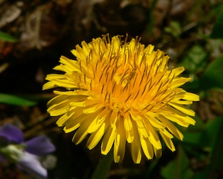
Scientific classification
Kingdom: Plantae
(unranked): Angiosperms
(unranked): Eudicots
(unranked): Asterids
Order: Asterales
Family: Asteraceae
Tribe: Cichorieae
Genus: Taraxacum
F. H. Wigg.
Type species
Taraxacum officinale [1]
F. H. Wigg.
Taraxacum /təˈræksəkʉm/ is a large genus of flowering plants in the family Asteraceae and consists of species commonly known as dandelion. They are native to Eurasia and North America, and two species, T. officinale and T. erythrospermum, are found as commonplace wild flowers worldwide.[2] Both species are edible in their entirety.[3] The common name dandelion (/ˈdændɨlaɪ.ən/ DAN-di-ly-ən, from French dent-de-lion, meaning "lion's tooth") is given to members of the genus. Like other members of the Asteraceae family, they have very small flowers collected together into a composite flower head. Each single flower in a head is called a floret. Many Taraxacum species produce seeds asexually by apomixis, where the seeds are produced without pollination, resulting in offspring that are genetically identical to the parent plant.[4]
http://yournewswire.com/weed-destroys-cancer/
University Study: Common Garden Weed Destroys Cancer
Cells In 48 Hours
by
Baxter Dmitry
by
Baxter Dmitry

Back in 2009, a group of Canadian researchers from the University of Windsor in Ontario started investigating a common weed as a potential cure for cancer, and the results are more than amazing.
It all started when an oncologist came across something quite interesting with some cancer patients. Believe it or not, the plant we are talking about is the common dandelion!
A post-doctoral fellow at the University of Windsor named Pamela Ovadje has done an extensive work on the topic. She dealt with the anti-cancer properties of dandelion and similar extracts.
According to Ovadje, “We had information from an oncologist, a collaborator here in Windsor, who had patients that showed improvement after taking dandelion root tea. And so, with a phone call, we decided to start studying what was in this tea that made patients respond to it, so we started digging up dandelions.”
She was quite suspicious in the beginning, but not because it was an all-natural source. “I figured dandelions are everywhere, and if there was something to it, people would have found this out already, “ she explained.
We should be glad to hear that the researchers have started conducting studies on dandelion root extract and its effects on cancer, as the results are astonishing.
“Since the commencement of this project, we have been able to successfully assess the effect of a simple water extract of dandelion root in various human cancer cell types in the lab and we have observed its effectiveness against human T-cell leukemia, chronic myelomonocytic leukemia, and pancreatic and colon cancers, with no toxicity to non-cancer cells. Furthermore, these efficacy studies have been confirmed in animal models (mice) that have been transplanted with human colon cancer cells.” [ http://www.uwindsor.ca/dandelionrootproject/ ]
Dandelion root extract was approved for human trials in February 2015. Now, it is in Phase 1 trials for end-stage blood-related cancers, such as leukemia and lymphoma.
According to Dr. Siyaram Pandey, professor of chemistry and biochemistry at the University of Windsor and principal research investigator for the project, dandelion root extract has quite a “good potential” to cause a death of cancer cells.
How Does it Work?
This extract causes cancer cells to go through apoptosis, a natural cell process where a cell activates an intracellular death program because it isn`t needed anymore. In brief, dandelion root extract causes the cancer cell to “commit suicide” without affecting the healthy ones.
Two cells perform apoptosis which is far better than chemotherapy drugs which kill one healthy cell for every 5 to 10 cancer cell, the dandelion extract.
It is important to mention that the concentration of this extract is much higher than the one which is currently available. Even though trials are still underway, this extract may be the future of cancer treatment!
The Dandelion Root Project is aimed at showcasing scientific evidence for the safe and effective use of dandelion root extract and other natural health products for cancer therapy.
The Dandelion Root Project started in 2009 in a bid to investigate the anticancer effect of the root extract of dandelions against cancer cells in the lab (in cells and in animal models). This project started with funding from the Knights of Columbus, Chapter 9671 (Windsor) and has been sustained by funding from other sources, including Seeds4Hope Grant (local Cancer Foundation), Lotte & John Hecht Foundation, The Pajama Angels and the Jesse & Julie Rasch Foundation. Private and personal donations have been made from the
Windsor local community, as well as from all around Canada. We dedicate this project in the memory of Mr. Kevin Couvillon, who lost his battle with leukemia in 2010.
Since the commencement of this project, we have been able to successfully assess the effect of a simple water extract of dandelion root in various human cancer cell types, in the lab and we have observed its effectiveness against human T cell leukemia, chronic myelomonocytic leukemia, pancreatic and colon cancers, with no toxicity to non-cancer cells. Furthermore, these efficacy studies have been confirmed in animal models (mice) that have been transplanted with human colon cancer cells.
We also applied for Phase I clinical trials in 2012 for the use of DRE in hematological cancers and in November 2012, we obtained approval for the administration of DRE in human patients and currently, the dandelion root extract is under Phase 1 clinical trials for drug refractory blood cancers.
Studies to understand how dandelion root extract can identify differences between cancer cells and non-cancer cells are underway, while at the same time, the identification of the active components within the extract is ongoing. We are excitingly awaiting the results from these studies.
US2016143979
Long Pepper Extract an Effective Anticancer Treatment
Long Pepper Extract an Effective Anticancer Treatment
Inventor(s): PANDEY SIYARAM, et al.
In a preferred embodiment, there is provided a method for preparing a medicament for the treatment or prevention of a cancer, the method comprising: grinding a Piper plant or a plant component thereof to obtain a ground plant mixture or powder; soaking the ground plant mixture or powder in a solvent to obtain a suspension having a liquid extract portion and a plant solid portion; and separating the liquid extract portion from the plant solid portion to provide a separated liquid extract for use in the medicament.
SCOPE OF THE INVENTION
[0002] The current invention relates to a method for preparing a medicament for the treatment or prevent of a cancer, and which broadly includes grinding a Piper plant or plant component to be extracted using a solvent, most preferably ethanol.
BACKGROUND OF THE INVENTION
[0003] The continuing increase in the incidence of cancer signifies a need for further research into more effective and less toxic alternatives to current treatments. In Canada alone, it was estimated that 267,700 new cases of cancer will arise, with 76,020 deaths occurring in 2012 alone. The global statistics are even more dire, with 12.7 million cancer cases and 7.6 million cancer deaths arising in 2008. The hallmarks of cancer cells uncover the difficulty in targeting cancer cells selectively. Cancer cells are notorious for sustaining proliferative signaling, evading growth suppression, activating invasion and metastasis and resisting cell death among other characteristics. These characteristics pose various challenges in the development of successful anticancer therapies. The ability of cancer cells to evade cell death events has been the center of attention of much research, with focus centered on targeting the various vulnerable aspects of cancer cells to induce different forms of Programmed Cell Death (PCD) in cancer cells, with no associated toxicities to non-cancerous cells.
[0004] Apoptosis (PCD type I) has been studied for decades, the understanding of which will enhance the possible development of more effective cancer therapies. This is a form of cell death that is required for regular cell development and homeostasis, as well as a defense mechanism to get rid of damaged cells; cells undergoing apoptosis invest energy in their own demise so as not to become a nuisance. Cancer cells evade apoptosis in order to confer added growth advantage and sustenance, therefore current anticancer therapies endeavor to exploit the various vulnerabilities of cancer cells in order to trigger the activation of apoptosis through either the extrinsic or intrinsic pathways. The challenges facing some of the available cancer therapies are their abilities to induce apoptosis in cancer cells by inducing genomic DNA damage. Although this is initially effective, as they target rapidly dividing cells, they are usually accompanied by severe side effects caused by the non-selective targeting of normal non-cancerous cells, suggesting a need for other non-common targets for apoptosis induction without the associated toxicities.
[0005] Currently chemotherapy is limited mostly to genotoxic drugs that are associated with severe side effects due to non-selective targeting of normal tissue. Natural products play a significant role in the development of most chemotherapeutic agents, with 74.8% of all available chemotherapy being derived from natural products. Natural health products (NHPs) have shown great promise in the field of cancer research. The past 70 years have introduced various natural products as the source of many drugs in cancer therapy.
Approximately 75% of the approved anticancer therapies have been derived from natural products, an expected statistic considering that more than 80% of the developing world's population is dependent on the natural products for therapy. Plant products especially contain many bioactive chemicals that are able to play specific roles in the treatment of various diseases. Considering the complex mixtures and pharmacological properties of many natural products, it becomes difficult to establish a specific target and mechanism of action of many NHPs. With NHPs gaining momentum, especially in the field of cancer research, there is a lot of new studies on the mechanistic efficacy and safety of NHPs as potential anticancer aunts. Long pepper, from the Piperaceae family, has been used for centuries for the treatment of various diseases. Several species of long pepper have been identified, including Piper Longum, Piper Belle, Piper Retrofactum, extracts of which have been used for years in the treatment of various diseases. A long list of uses and benefits are associated with extracts of different piper spp, with reports indicating their effectiveness as good digestive agents and pain and inflammatory suppressants. However, there is little to no scientific validation, only anecdotal evidence, for the benefits associated with the use of long pepper extracts. There are scientific studies have been carried out on several compounds present in extracts of long pepper, including piperines, which has been shown to inhibit many enzymatic drug bio-transforming reactions and plays specific roles in metabolic activation of carcinogens and mitochondrial energy production, and various piperidine alkaloids, with fungicidal activity.
SUMMARY OF INVENTION
[0006] One possible non-limiting object of the present invention is to provide a method for preparing a medicament for the treatment or prevention of a cancer, and which includes as a main active ingredient substances derived from a readily available natural health product.
[0007] Another possible non-limiting object of the present invention is to provide a method for preparing a cancer medicament which does not strictly require inclusion of synthetic genotoxic drugs often associated with undesirable side effects and non-selective targeting of both cancerous and non-cancerous cells.
[0008] Another possible non-limiting object of the present invention is to provide a method for preparing a cancer medicament which may permit for more selective treatment of cancer cells, while reducing production costs.
[0009] In one aspect, the present invention provides a method for preparing a medicament for the treatment or prevention of a cancer, the method comprising: grinding a Piper plant or a plant component thereof to obtain a ground plant mixture or powder; soaking the ground plant mixture or powder in a solvent to obtain a suspension having a liquid extract portion and a plant solid portion; and separating the liquid extract portion from the plant solid portion to provide a separated liquid extract for use in the medicament.
[0010] In another aspect, the present invention provides a method for preparing a medicament comprising a Piper plant extract for treatment or prevention of a cancer, the method comprising the steps of: grinding a Piper plant seed to obtain a ground seed powder; steeping the ground seed powder in a solvent comprising ethanol to obtain a mixture having a liquid extract portion and a solid portion; separating the liquid extract portion from the solid portion, and removing the solvent from the liquid extract portion to obtain a solid or semi-solid extract; and optionally mixing the solid or semi-solid extract with a polar reconstitution solvent comprising dimethyl sulfoxide, water, alcohol or a mixture thereof, said alcohol preferably comprising one or more of n-butanol, isopropanol, n-propanol, ethanol and methanol.
[0011] In yet another aspect, the present invention provides a method of treating or preventing a cancer, the method comprising administering to a subject an effective amount of a medicament prepared by the method of the present invention.
[0012] To scientifically assess the anticancer potential of a preferred ethanolic extract of Long pepper (hereinafter also referred to as “PLX”), a plant of the Piperaceae family was selected with a view to assessing the efficacy of the anticancer mechanism of action of PLX against cancer cells. It has been recognized that the preferred ethanolic long pepper extract selectively induce caspase-independent apoptosis in cancer cells, without affecting non-cancerous cells, by targeting the mitochondria, leading to dissipation of the mitochondrial membrane potential and increase in reactive oxygen species or ROS production. Release of the AIF and endonuclease G from isolated mitochondria confirmed the mitochondria as a potential target of long pepper. The efficacy of PLX in in vivo studies indicates that oral administration may slow or even be able to halt the growth of colon cancer tumors in immunocompromised mice, with no associated toxicity. These results demonstrate the potentially safe and non-toxic alternative that is long pepper extract for cancer therapy.
[0013] It is to be appreciated that the Piper plant in its entirety or one or more plant components thereof may be utilized for preparing a medicament for treatment or prevention of a cancer. In one embodiment, the plant component comprises one or more of a seed, a leaf, a flower, a fruit, a root and a stem, or more preferably a seed.
[0014] In one embodiment, the solvent comprises one or more of water, pentane, cyclopentane, cyclohexane, benzene, toluene, 1,4-dioxane, chloroform, diethyl ether, dichloromethane, tetrahydrofuran, ethyl acetate, acetone, dimethylformamide, acetonitrile, dimethyl sulfoxide, propylene carbonate, formic acid, alcohol and acetic acid, wherein the alcohol preferably includes one or more of n-butanol, isopropanol, n-propanol, ethanol and methanol. In one embodiment, the solvent is ethanol.
[0015] In one embodiment, said soaking or steeping step comprises soaking or steeping the ground plant mixture or powder with or without shaking in the solvent for between about 5 minutes and about 72 hours, preferably between about 4 hours and about 48 hours, more preferably between about 12 hours and about 36 hours, or most preferably for about 24 hours, and at a temperature between about 0° C. and about 100° C., preferably between about 10° C. and about 75° C., more preferably between about 15° C. and about 50° C., or
most preferably about 25° C.
[0016] It is contemplated further process improvements in the extraction method may be implemented. These may, for example, include: cryogenic grinding of the plant material to for example micronized or nano scale particles; harvesting the long pepper fruit when the active compounds are at their highest concentrations; and better storage of long pepper between harvest and process to preserve the active compounds at their optimum levels.
[0017] In one embodiment, the method further comprises freezing the Piper plant or the plant component to obtain a frozen plant stock, said freezing step being selected to effect at least partial disruption of one or more plant cells, and wherein said grinding step comprises dry grinding the frozen plant stock to obtain the ground plant powder, wherein during said dry grinding step, the frozen plant stock is maintained at a grinding temperature below about 40° C., preferably below about 0° C., more preferably below about −25° C. or most preferably below about −40° C. Preferably, said freezing step comprises contacting or submerging the Piper plant or the plant component in liquid nitrogen, or freezing the Piper plant or the plant component to an average freezing temperature between about −210° C. and about −30° C. In one embodiment, said dry grinding step comprises dry grinding the frozen plant stock to an average particle size of less than about 100 μm, preferably less than about 70 μm or more preferably less than about 45 μm.
[0018] In one embodiment, prior to said freezing step, the method further comprises drying said Piper plant or the plant component to a relative humidity between about 5% and about 10%.
[0019] In one embodiment, said dry grinding step comprises dry grinding the frozen plant stock with a grinder selected from the group consisting of a pulverizer, an impingement grinder and a micronized milling machine, and wherein the grinder or a component thereof is cooled below about −25° C. or preferably below about −50° C., to prevent heating on contact with the frozen plant stock or the ground plant powder. In one embodiment, the grinder or a component thereof is cooled by directly or indirectly contacting with liquid nitrogen. In one embodiment, the grinder defines a grinding chamber sized for receiving the frozen plant stock, said method further comprising flowing or adding liquid nitrogen to the grinding chamber during said dry grinding the frozen plant stock in the grinding chamber. In one embodiment, said dry grinding step further comprises straining the ground plant powder from the grinder through a sieve sized to obtain a sieved ground plant powder having an average particle size of less than about 100 μm, preferably less than about 70 μm or more preferably less than about 45 μm. The applicant has appreciated that such dry grinding to obtain the sieved ground plant powder may permit for improved extraction of active ingredients from the Piper plant, while reducing loss of bioactivity.
[0020] In one embodiment, said separation step comprises of filtration, wherein said filtration is performed once or more than once using a plurality of filters of same or different pore sizes. Preferably, said separation step comprises filtering the suspension at least twice with a paper filter having a particle retention greater than about 20 μm.
[0021] It is to be appreciated that the liquid extract portion may be subject to further processing. In one embodiment, said method further comprises removing the solvent from the separated liquid extract to obtain a solid or semi-solid extract, and optionally mixing the extract with one or more of a pharmaceutically acceptable carrier, a diluent, a binding agent, an adjuvant and an anticancer aunt. The solid or semi-solid extract may be administered to a subject. In one embodiment, the carrier comprises one or more of a polar reconstitution solvent and a buffer solution, the reconstitution solvent preferably comprising dimethyl sulfoxide, alcohol or a mixture thereof, the alcohol preferably comprising one or more of n-butanol, isopropanol, n-propanol, ethanol and methanol, and the buffer solution preferably comprising a phosphate buffered saline solution or a sodium bicarbonate buffered saline solution. In one embodiment, the anticancer agent comprises metformin, hydroxyurea, cyclophosphamide, etoposide or another anticancer natural extract.
[0022] It is to be appreciated that the Piper plant species for use with the current invention is not intended to be specifically limited to Piper longum, and may alternatively include among others Piper belle, Piper retrofactum and Piper nigrum. Indeed, the applicant has appreciated that the method may be practiced with a Piper plant other than Piper longum, while retaining anticancer activity of the medicament. Preferably, the Piper plant is selected to provide in the liquid extract portion or the medicament two or more of dihydropiperlongumine, piperlongumine, dihydropiperlonguminine, piperlonguminine, piperinine, piperazine, piperidine and piperine, or preferably two or more of dihydropiperlongumine, piperlongumine, dihydropiperlonguminine, piperlonguminine, piperinine and piperine. The applicant has appreciated that the aforementioned compounds may provide for anticancer activities.
[0023] The medicament of the current invention may permit for treatment or prevention of a cancer, including but not limited to colorectal cancer, ovarian cancer, pancreatic cancer, melanoma, breast cancer, osteosarcoma, lung cancer, prostate cancer, glioblastoma, lymphoma or leukemia, or preferably colorectal cancer, ovarian cancer, pancreatic cancer, melanoma, glioblastoma or leukemia, wherein the leukemia, includes T cell leukemia, acute myeloid leukemia, chronic myeloid leukemia, chronic myelomonocytic leukaemia, chronic lymphocytic leukemia or acute lymphoblastic leukemia. Some clinical studies have been conducted to evaluate anticancer potential or activities of preferred medicaments of the present invention, including those for glioblastoma.
[0024] In one embodiment, the method further comprises freezing the Piper plant seed to obtain a frozen seed stock, and wherein said grinding step comprises dry grinding the frozen seed stock to obtain the ground seed powder, wherein during said dry grinding step, the frozen seed stock is maintained at a grinding temperature below about 0° C., preferably below about −25° C. or more preferably below about −40° C. Preferably, said freezing step comprises contacting or submerging the Piper plant seed in liquid nitrogen, or
freezing the Piper plant seed to an average freezing temperature between about −210° C. and about −30° C. Preferably, said dry grinding step comprises dry grinding the frozen seed stock to an average particle size of less than about 100 μm. preferably less than about 70 μm or more preferably less than about 45 μm with a grinder selected from the group consisting of a pulverizer, an impingement grinder and a micronized milling machine, wherein the grinder or a component thereof is cooled below about −25° C. or preferably below about −50° C., to prevent heating on contact with the frozen seed stock or the ground seed powder.
[0025] In one embodiment, the grinder or a component thereof is cooled by directly or indirectly contacting with liquid nitrogen. In one embodiment, the grinder defines a grinding chamber sized for receiving the frozen plant stock, said method further comprising flowing or adding liquid nitrogen to the grinding chamber during said dry grinding the frozen plant stock in the grinding chamber. In one embodiment, said dry grinding step further comprises straining the ground plant powder from the grinder through a sieve sized to obtain a sieved ground plant powder having an average particle size of less than about 100 μm, preferably less than about 70 μm or more preferably less than about 45 μm. The applicant has appreciated that such dry grinding to obtain the sieved ground plant powder may permit for improved extraction of active ingredients from the Piper plant, while reducing loss of bioactivity.
[0026] In one embodiment, said mixing step comprises mixing the solid or semi-solid extract with the polar reconstitution solvent to obtain a reconstituted extract, and mixing the reconstituted extract with a buffer solution and optionally an anticancer agent, the buffer solution preferably comprising a phosphate buffered saline solution or a sodium bicarbonate buffered saline solution, and the anticancer agent preferably comprising metformin, hydroxyurea, cyclophosphamide or etoposide.
BRIEF DESCRIPTION OF THE DRAWINGS
[0027] Reference may now be had to the following detailed description taken together with the accompanying drawings which:
[0028] FIG. 1 shows four (4) bar graphs illustrating absorbance values at 450 nm expressed as a percent of the control for colon (HCT116), ovarian (OVCAR-3), pancreatic (BxPC-3) cancer and Melanoma (G-361) cells treated with a crude ethanolic extract of long pepper at indicated concentrations, and then incubated with WST-1 cell viability dye for 4 hours. The values are expressed as mean±SD from quadruplicates of 3 independent experiments, with **P<0.0001;
[0029] FIG. 2 shows ten (15) bar graphs illustrating the results of image-based cytometry for induction of cell death in respect of human pancreatic (BxPc-3) cancer and T cell leukemia cells treated with PLX at indicated concentrations, and which were subsequently incubated with propidium iodide at indicated time points;
[0030] FIG. 3 shows six (6) images of human colon cancer cells (HT-29) treated with PLX at indicated concentrations, and subsequently incubated with propidium iodide, and which are obtained from fluorescence microscopy (at 400× magnification on a fluorescent microscope, scale bar=15 μm) for assessing induction of cell death as characterized by presence of propidium iodide positive cells;
[0031] FIG. 4 shows six (6) images of normal colon epithelial cells (NCM460) treated with PLX at indicated concentrations, and subsequently incubated with propidium iodide, and which are obtained from fluorescence microscopy (at 400× magnification on a fluorescent microscope, scale bar=15 μm) for assessing induction of cell death as characterized by presence of propidium iodide positive cells;
[0032] FIG. 5 shows nineteen (19) bar graphs illustrating the results of image-based cytometry for apoptotic induction (% annexin V positive) and necrosis (% PI positive) in respect of E6-1 cells following PLX treatment at indicated concentrations, with the lack of annexin V or PI staining indicating live cells following the treatment (% annexin V/PI negative cells, *P<0.05, **P<0.003, ***P<0.0001);
[0033] FIG. 6 shows nineteen (19) bar graphs illustrating the results of image-based cytometry for apoptotic induction (% annexin V positive) and necrosis (% PI positive) in respect of HT-29 cells following PLX treatment at indicated concentrations, with the lack of annexin V or PI staining indicating live cells following the treatment (% annexin V/PI negative cells, *P<0.05, **P<0.003, ***P<0.0001);
[0034] FIG. 7 shows eight (8) bar graphs illustrating the results of image-based cytometry for detection of DNA fragmentation with TUNEL labeling in respect of cancer cells treated with PLX and VP16 (as a positive control for DNA damage), and which were subsequently labelled with DNA staining solution and quantified by image-based cytometry (treated cells were compared to the control untreated cell sample, ***P<0.0001);
[0035] FIG. 8 shows eighteen (18) images of OVCAR-3, G-361 and NCM460 cells obtained with a fluorescent microscope at 400× magnification (scale bar=15 μm), and which were treated with PLX at indicated concentrations and stained with Hoechst to characterize nuclear morphology and Annexin-V to detect apoptotic cells;
[0036] FIG. 9 shows eight (8) images of HT-29 and NCM460 cells obtained with a fluorescent microscope at 400× magnification (scale bar=15 μm), and which were treated with PLX at indicated concentrations and stained with Hoechst to characterize nuclear morphology and subject to phase contrast microscopy for cellular morphology;
[0037] FIG. 10 shows two (2) bar graphs illustrating percent viability values for HT-29 colorectal cancer cells and non-cancerous NCM460 cells treated with PLX and subsequently incubated with WST-1 cell viability dye for 4 hours, and which were obtained by measuring absorbance values at 450 nm and expressed as a percent of the control (values are expressed as mean±SD from quadruplicates of 3 independent experiments. **P<0.0001);
[0038] FIG. 11 shows a bar graph illustrating fluorescent readings (an average of 6 readings per well and a minimum of three wells per experiment, and the average of three independent experiments shown) expressed as activity per μg of protein (in fold), and which are obtained with a spectrofluorometer in respect of cell lysate of BxPc-3 cells incubated with caspase substrates specific to each caspase 3, 8 or 9 for an hour, where the BxPc-3 cells were treated with 0.10 mg/ml PLX at indicated time points, collected, washed
and incubated with lysis buffer;
[0039] FIG. 12 shows two (2) bar graphs illustrating absorbance values at 450 nm expressed as a percent of the control in respect of HCT 116 and BxPC-3 cancer cells pretreated or not pretreated with Z-VAD-fink for an hour before PLX treatment at indicated concentrations, and which are representative of a WST-1 cell viability assay of the cancer cells (the values are expressed as mean±SD from quadruplicates of 3 independent experiments. **P<0.0001);
[0040] FIG. 13 shows nine (9) bar graphs illustrating fluorescence results obtained with an image based cytometer in respect of colon cancer (HT-29), normal colon epithelial (NCM460) and normal human fibroblast (NHF) cells treated with PLX at indicated concentrations for 48 hours, and subsequently treated with H2DCFDA;
[0041] FIG. 14 shows a bar graph illustrating quantified results of the fluorescence results shown in FIG. 13 using Graphpad prism
6.0;
[0042] FIG. 15 shows a bar graph illustrating absorbance values at 450 nm expressed as a percent of the control in respect of HCT 116 colon cancer cells treated with 3 mM N-acetylcysteine for an hour, then with PLX at indicated concentrations for 72 hours and being subject a WST-1 assay (the values are expressed as mean±SD from quadruplicates of 3 independent experiments. **P<0.05);
[0043] FIG. 16 shows eight (8) bar graphs illustrating fluorescence values obtained using image based cytometry in respect of colon cancer (HT-29), ovarian cancer (OVCAR-3) and normal colon epithelial (NCM460) cells treated with PLX at indicated concentrations for 48 hours, and subsequently incubated with JC-1;
[0044] FIG. 17 shows a bar graph illustrating quantified results of the fluorescence results shown in FIG. 16;
[0045] FIG. 18 shows eight (8) images of OVCAR-3 and NCM460 cells taken at 400× magnification using a fluorescent microscope (scale bar=15 μm), and which were treated with PLX, and subsequently incubated with TMRM cationic mitochondrial membrane permeable dye (corresponding Hoechst dye images are also shown);
[0046] FIG. 19 shows on the left hand portion a series of images obtained from western blot analyses for pro-apoptotic factors AIF and EndoG in mitochondrial supernatants obtained by centrifuging samples of isolated mitochondria of OVCAR-3 cells treated directly with PLX or solvent control (ethanol) for 2 hours (the mitochondrial pellets were probed for SDHA to serve as loading controls, and the images are representative of 3 independent experiments demonstrating similar trends), and on the left hand portion two (2) bar graphs showing ratios of AIF or EndoG to SDHA (the values are expressed as mean±SD of quadruplicates of 1 independent experiment; *p<0.01 versus solvent control (ethanol));
[0047] FIG. 20 shows a bar graph (left) and a table (right) illustrating results from a protein urinalysis by Bradford assay and dipstick analysis conducted in respect of BALB/C mice divided into three groups, or namely a control group (3 animals, untreated and given plain filtered water), a gavage control group (3 animals, given 50 mg/kg/day vehicle (DMSO) and a treatment group (4 animals, given 50 mg/kg/day PLX);
[0048] FIG. 21 shows a line graph illustrating weight changes of the BALB/c mice detailed above in respect of FIG. 20;
[0049] FIG. 22 shows six images of hematoxylin and eosin stained tissue sections of the liver, heart and kidney of the BALB/c mice detailed above in respect of FIG. 20, and which were obtained on a bright field microscope at 63× objective;
[0050] FIG. 23 shows nine (9) photographs illustrating representative tumor sizes on CD-1 nu/nu mice subcutaneously injected with colon cancer cells (HT-29 (p53<−/−>) on the left flank and HCT116 (p53<+/+>) on the right flank), and which were divided into three groups, or namely a control group, a gavage control group and a treatment group;
[0051] FIG. 24 shows two (2) line graphs illustrating average body weights and tumor volumes of the CD-1 nu/nu mice detailed above in respect of FIG. 23 over time;
[0052] FIG. 25 shows twenty (20) images illustrating histopathological analysis of tissue samples obtained from the CD-1 nu/nu mice detailed above in respect of FIG. 23, and which represents hematoxylin and eosin stained tissue sections of the livers, hearts, kidneys and tumors (the images were obtained on a bright field microscope at 10× and 63× objective); and
[0053] FIG. 26 shows two (2) chromatograms of a piperamides standard mix (1 mg/mL at 1 μL/standard) and a PLX (10 mg/mL at 2 μL/sample).
DETAILED DESCRIPTION OF THE PREFERRED EMBODIMENTS
[0054] Indian long pepper seeds were obtained from Quality Natural Foods limited, Toronto Ontario. The plant material was ground up and extracted in anhydrous ethanol (100%) in a ratio of about 1:10. The extraction was carried out overnight on a shaker at room temperature. The extract was passed through a P8 coarse filter, followed by a 0.45 μn1 filter. The solvent was evaporated using a rotary evaporator at 40° C. resulting in a residual powdered concentrate. This powder concentrate was then reconstituted in
dimethylsulfoxide (Me2SO) at a stock concentration of approximately 450 mg/ml. When administered the reconstituted solution is mixed with phosphate buffered saline solution and given orally. The applicant has recognized that Long pepper extracts may represent a new NHP, with better selective efficacy against cancer cells.
[0055] Alternatively, a preferred long pepper extract may be prepared with whole Piper longum or seeds thereof purchased from Premier Herbal Inc. of Toronto, Ontario, and which originate from India.
[0056] The applicant has examined the efficacy of an ethano lie extract of Long Pepper against various cancer cells, and has attempted to elucidate the mechanism of action, following treatment. Results from this preliminary studies suggest that PLX may reduce the viability of various cancer cell types in a dose and time dependent manner, where apoptosis induction was observed, following mitochondrial targeting. Due to the low doses of PLX required to induce apoptosis in cancer cell a therapeutic window of
this extract is furthermore suggested. Preliminary studies suggest the induction of apoptosis may be caspase-independent, although there was activation of both the extrinsic and intrinsic pathways and the production of ROS was not essential to the mechanism of cell death induction by PLX. The ability of PLX to target multiple vulnerabilities of cancer cells and still act to induce apoptosis in the presence of different types of inhibitors suggests the potential application of PLX in safe and efficacious cancer therapy.
[0057] In one experiment, following treatment with ethanolic long pepper extract, cell viability was assessed using a water-soluble tetrazolium salt; apoptosis induction was observed following nuclear staining by Hoechst, binding of annexin V to the externalized phosphatidyl serine and phase contrast microscopy. Image-based cytometry was used to detect the effect of long pepper extract on the production of reactive oxygen species and the dissipation of the mitochondrial membrane potential following Tetramethylrhodamine or
5,5,6,6′-tetrachloro-1,1′,3,3′-tetraethylbenzimidazolylcarbocyanine chloride staining (JC-1). Assessment of PLX in vivo was carried out using Balb/C mice (toxicity) and CD-1 nu/nu immunocompromised mice (efficacy). HPLC analysis enabled detection of some primary compounds present within our long pepper extract. Without being bound by a particular theory, preliminary testing results suggest that an ethanolic long pepper extract may selectively induce caspase-independent apoptosis in cancer cells, without affecting non-cancerous cells, by targeting the mitochondria, leading to dissipation of the mitochondrial membrane potential and increase in ROS production. Release of the AIF and endonuclease G from isolated mitochondria confirms the mitochondria as a potential target of long pepper. The efficacy of PLX in in vivo studies indicates that oral administration may be able to retard or halt the growth of colon cancer tumors in immunocompromised mice, with no associated toxicity. These results suggest the potentially safe and non-toxic alternative that is long pepper extract for cancer therapy.
[0058] To further explicitly illustrate the effectiveness of the medicament of the present invention, detailed descriptions of exemplary experiments are provided below:
i) Ethanolic Extract of Long Pepper Effectively and Selectively Reduces the Viability of & Induces Apoptosis in Cancer Cells in a Dose & Time Dependent Manner
[0059] In one experiment, as a first step in assessing the effect of long pepper extract the effect of PLX on the viability of cancer cells was assessed. In particular, following treatment with increasing concentration of PLX at increasing time points, cells were incubated with a water soluble tetrazolium salt, which gets metabolized to a red formazan product by viable cells with active metabolism. This product can then be quantified by absorbance spectrometry. The efficacy of crude PLX in reducing the viability of cancer cells was observed, including colon (HCT116), pancreatic (BxPC-3), ovarian cancer (OVCAR-3) and melanoma cells. This effect was dose and time dependent (see FIG. 1).
[0060] To further evaluate the anticancer activity of PLX, its role in cell death and its selectivity to cancer cells was assessed. The preliminary results demonstrate that PLX may be able to selectively induce cell death in cancer cells (colon, pancreatic and leukemia) in a dose and time dependent manner, as characterized by the increase in propidium iodide positive cells in cancer cells treated with PLX (see FIGS. 2 and 3). Furthermore, this effect was selective, as normal colon epithelial cells remained substantially unaffected by this treatment, at the same concentrations and time-points (see FIGS. 3 and 4). Additionally, apoptosis induction in various cancer cells, melanoma (G-361), ovarian and colon cancer (HT-29) cells, was confirmed by Annexin-V binding assay. This induction of apoptosis was confirmed to be selective to cancer cells, as normal colon cells (NCM460) remained unaffected by PLX treatment. This was indicated by nuclear condensation, cell morphology and externalization of phosphatidyl serine to the outer leaflet of the cell membrane, as indicated by Hoechst staining, phase contrast images and binding of annexin V dye respectively (see FIGS. 8 to 10). The selectivity of PLX to cancer cells was further confirmed by the WST-1 cell viability assay that showed that PLX was highly effective at such low doses, a therapeutic window was easily observed (see FIG. 10). Treatment of HT-29 with 0.20 mg/ml effectively reduced the viability by approximately 90%, while NCM460 cells remained at 100% viability at the same dose. This indicates that PLX can be more effective at very low doses, further reducing the chances of toxicity associated with treatment.
[0061] To confirm the induction of apoptosis, the binding of Annexin V to externalized phosphatidylserine on the outer cellularsurface, was assessed. Following treatment with PLX, cells (FIG. 5—E6-1 and FIG. 6—HT-29 cells) were washed twice in phosphate buffer saline (PBS). Subsequently, cells were resuspended and incubated in Annexin V binding buffer (10 mM HEPES, 10 mM NaOH, 140 mM NaCl, 1 mM CaCl2, pH 7.6) with Annexin V Alexa Fluor-488 (1:50) (Invitrogen, Canada, Cat No. A13201) for 15 minutes. In the final 10 minutes of incubation, 1 mg/ml propidium iodide was added to the microcentrifuge tube and incubated for the final 10 minutes in the dark. Image based cytometry was used to quantify the percentage of programmed cell death (annexin V positive cells) and necrotic cell death (propidium iodide positive cells) occurring after treatment (see FIGS. 5 and 6)
[0062] Following PLX treatment, HT-29 cells were labeled with the Terminal deoxynucleotidyl transferase dUTP nick end labeling (TUNEL) assay. The assay was performed according to the manufacturer's protocol (Molecular Probes, Eugene, Oreg.), in order to detectDNA damage Cells were treated with PLX or VP-16 (as a positive control) at indicated concentrations and time points and analyzed for the fragmentation of DNA. Following treatment, cells were fixed by suspending them in 70% (v/v) ethanol and stored at −20° C.
overnight. The sample was then incubated with a DNA labeling solution (10 μL reaction buffer, 0.75 μL TdT enzyme, 8 μL BrdUTP, 31.25 μL of dH2O) for 1 hour at 25° C. Each sample was exposed to an antibody solution (5 μL Alexa Fluor 488 labeled anti-BrdU antibody and 95 μL rinse solution). The cells were incubated with the antibody solution for 20 minutes and TUNEL positive cells were quantified by image-based cytometry (see FIG. 7).
ii) PLX Induces Caspase-Independent Apoptosis in Human Cancer Cells
[0063] It has been recognized that caspases are cysteine aspartic proteases that play a predominant role as death proteases. Their roles in various cell death processes remains controversial, as their activation or inhibition could be essential to the progression of inhibition of cell death pathways. In another experiment, to assess the role of caspases, following treatment with 0.10 mg/ml PLX, at indicated time points, BxPc-3 cells were collected, washed and incubated with lysis buffer to obtain cell lysate. The cell lysate
was incubated with caspase substrates, specific to each caspase (3, 8 and 9) and incubated for an hour. Fluorescence readings were obtained using a spectrofluorometer. Results indicate that PLX is able to activate both pathways (extrinsic and intrinsic apoptosis) in a time dependent manner. This was observed as rapid activation of caspases-3, 8 and 9 were observed as early as an hour, following treatment (see FIG. 11). To determine the importance of these activated caspases to the apoptosis-inducing effect of PLX, colon (HCT116) and pancreatic (BxPc-3) cancer cells were pre-treated with a pan-caspase inhibitor, Z-VAD-fink (20 μM), for an hour before treatment with PLX. Following treatments, the VST-1 cell viability assay was used to assess for viability and efficacy of PLX. Our results indicate that the inhibition of caspases may not prevent the reduction of viability (see FIG. 12), suggesting that the effect of PLX in cancer cells may be caspase independent.
iii) Long Pepper Extract Induces Oxidative Stress and Targets the Mitochondria of Cancer Cells
[0064] Generation of oxidative stress has been well established as a major player in the induction of several cell death processes, especially apoptosis. In another experiment, the applicant has also examined the role of oxidative stress in PLX induced apoptosis. Following treatment with PLX for 48 hours, cells were incubated with 2′,7′-Dichlorofluorescin diacetate H2DCFDA for 45 minutes. The resulting green fluorescence histograms were obtained using a TALI image-based cytometer.
[0065] From the results, it was observed that PLX induced extensive generation of whole cell reactive oxygen species (ROS) in HT-29 colon cancer cells, while acting to suppress any ROS present in the non-cancerous cell lines, NCM460 and normal human fibroblasts (NHF) (see FIGS. 13 and 14). This appears to confirm the results of selectivity and indicates that PLX might act as a pro-oxidant in cancer cells in order to induce apoptosis.
[0066] To determine if this oxidative stress was essential to PLX activity, HCT116 colon cancer cells were pre-treated with N-acetyl-L-cysteine (NAC), a well-established anti-oxidant, used extensively in vitro studies, before treatment with PLX. Subsequent to PLX treatment, cells were analyzed for effect of PLX on viability, using the WST-1 viability assay. The results suggest that although PLX may act to induce oxidative stress to cause apoptosis, this oxidative stress is not essential to its activity. Both the cells
treated with PLX alone and NAC followed by PLX showed a reduction in their viability (see FIG. 15).
[0067] The mitochondria have also been shown to play a major role in the progression and execution of apoptosis. The permeabilization of the mitochondrial membrane usually leads to the release of pro-apoptotic factors, including cytochrome c, apoptosis inducing factor (AIF) and endonuclease G (EndoG). These factors cause a caspase-independent pathway for apoptosis to pass through and could bypass the antioxidant effects of NAC observed in FIG. 15.
[0068] In another experiment, to assess the efficacy of PLX on the mitochondria of cancer cells, OVCAR-3, HT-29 and NCM460 cells were stained with TMRM, a cationic dye that accumulates in healthy mitochondria. Mitochondrial membrane potential (MMP) dissipation was only observed in OVCAR-3 and HT-29 cells as seen with the dissipation of red TMRM fluorescence, by fluorescence microscopy and image-based cytometry (see FIGS. 16 to 18). Following mitochondrial membrane collapse, we wanted to determine if there was release of some pro-apoptotic factors. Western blot analysis was used to monitor for the release of AIF and EndoG from isolated OVCAR-3 mitochondria. Results demonstrate that PLX directly caused the release of both AIF and EndoG from the mitochondria of OVCAR-3 cells (see FIG. 19). These results provide an insight to the mechanism of PLX action, where the mitochondria appears to be a direct target of PLX for the reduction of viability and the induction of apoptosis.
iv) Long Pepper Extract is Well-Tolerated in Animal Models
[0069] In another experiment, to scientifically evaluate and validate the safety of PLX, balb/c mice were orally gavaged with 50 mg/kg/day vehicle (DMSO) or PLX for 75 days and the mice were observed for signs of toxicity. To assess for toxicity, mice were weighed twice a week, urine was collected for protein urinalysis studies and following period of treatment, mice were sacrificed and their organs were obtained for pathological analysis by a certified pathologist at the University of Guelph (Dr. Brookes). Results from this part of the study demonstrate that there was no weight loss overall in mice that were given PLX supplemented water (see FIG. 21). To further assess toxicity, urine was collected from mice once a week and protein urinalysis was performed using a urine dipstick and a Bradford protein concentration assay. Protein urinalysis results indicate that there were trace amounts of protein in the urine of mice both from the control and the PLX group, with trace readings corresponding to protein concentrations between 5 and 20 mg/dL (data not shown). Bradford assays confirm the results obtained by dipstick urinalysis (see FIG. 20). There was no major difference between the control group and PLX group, confirming the lack of toxicity associated with oral administration of PLX in drinking water. Furthermore, the hearts, livers and kidneys were obtained following the toxicity study, sliced and stained with hematoxylin and eosin. Results show no gross morphologic difference between the control and the treatment group, confirming the lack of toxicity associated with PLX treatment. Results from the pathologist, indicate that the presence of any lesions in the tissues are minimal or mild and interpreted as either background or incidental lesions, and the lack of lesion type and frequency was enough to conclude no toxicological effect of PLX to the balb/c mice (Table 1).
TABLE 1
Summary of Histological Lesions in Balb/C Mice on PLX regimen
No Vehicle Long
Treat- (Gavage Pepper Extract
ment Control) (Treatment group)
M1 M2 M1 M2 M3 M1 M2 M3 M4
Liver:
Infiltration, leukocyte, X X X X X
predominantlymononuclear, minimal
Focal mineralization, minimal
Hepatocyte necrosis, minimal X
Focus of cellular alteration, X X X
eosinophilic, minimal Hepatocyte vacuolation, X X X
lipid type, minimal
Hepatocyte vacuolation, X X X X
lipid type, mild
Fibrin thrombus X
Heart:
Infiltration, leukocyte, X X X
predominantlymononuclear, minimal
Myofiber separation X X X
and vaculation,minimal (suspect artifact)
Kidney:
Infiltration, leukocyte, X X X X X
predominantly mononuclear,minimal
Tubule vacuolation, minimal X X
Fibrin or other extracellular X
matrix, glomerulus
v) Oral Administration of Long Pepper Extract Halts the Growth of Human Colon Cancer Xenografts in Immunocompromised Mice
[0070] In another experiment to study the efficacy of PLX, CD-1nu/nu immunocompromised mice were subcutaneously injected with HT-29 cells (left) and HCT116 cells (right). Following the establishment of tumors, mice were separated into three groups, a control group, a vehicle (Me2SO) group and a PLX treated group. Mice were observed for 75 days, with weights and tumor volumes measured twice a week. Results demonstrate that oral administration of PLX could suppress the growth of both p53 WT (HCT116) and p53 mutant (HT-29) tumors in-vivo. There were no signs of toxicity, as indicated by increasing weights during the study (see FIGS. 23 and 24). Furthermore, H & E staining revealed less nuclei in the PLX treated group, compared to the control group, however, as observed in the toxicity studies, there were no gross morphological differences in the livers, kidneys and hearts of the control and PLX groups (see FIG. 25).
vi) Analysis of Long Pepper Extract
[0071] Our collaborators at the University of Ottawa ran an HPLC profile study on the crude ethanolic extracts, compared with a piperamide standard mix. The chromatogram profile showed that our PLX extract contained several classes of compounds known to be present in piper species, including piperines and dihydropiperlongumine. We observed a lack of piperlongumine peaks in our PLX extract (see FIG. 26), suggesting that the PLX may be obtained from the species Piper Belle or the Piper Retrofactum.
[0072] The applicant has appreciated the selective anticancer potential of an ethano lie extract of long pepper in several cancer and non-cancerous counterparts. The preferred PLX was shown to effectively reduce the viability of cancer cells, and induced apoptosis in a dose- and time-dependent manner, at low doses, allowing for a greater therapeutic window in in-vitro studies (see FIGS. 1 to 4 and 8 to 10). This apoptosis inducing effect was found to be independent of caspases, cysteine aspartic proteases that play a role in the
progression and execution of apoptosis (see FIG. 12). These results suggest that PLX is not toxic to non-cancerous cells at such low doses, as was observed in the cancer cells. Selectivity and lack of toxicity was shown with in-vivo toxicological studies. Furthermore, damage to the kidneys is a common occurrence during various types to toxic therapies. This damage to the kidney results in large amounts of protein (>3.5 g/day) leaking into the urine, and this can be measured by various assays. Lack of toxicity was shown by the lack of increased protein concentration in the urine samples collected from both the control group and PLX treated group, by two different assays. The urine dipstick method indicated that all urine samples from the control and PLX groups had trace amounts of protein, corresponding to concentrations between 5 mg/dL and 20 mg/dL, well within the acceptable concentration range. Bradford protein assay showed a concentration of approximately 30 mg/dL most days urine was collected (see FIG. 20). This is still within the acceptable range of protein concentration in urine. These results suggest reduced associated toxicity or side effects observed with take long pepper extracts. The efficacy of PLX in in-vivo models also showed that not only was PLX well-tolerated, it was also effective at halting the growth of human tumor xenografts of colon cancer in nude mice (see FIGS. 20 and 21).
[0073] The next step in understanding the effect of PLX on cell death induction in cancer cells was to identify the mechanism of apoptosis induction observed following PLX treatment. The role of oxidative stress in cell death processes has been well characterized. It is well established the reactive oxygen species (ROS) could be the cause or effect of apoptosis induction in cells. Some studies have suggested cancer cells to be more dependent on cellular response mechanisms against oxidative stress and have exploited this feature to selectively target cancer cells. The role of ROS generation in PLX-induced apoptosis was assessed following treatment. In this study, we found that PLX induced whole cell ROS production in a dose dependent manner, as indicated by the increase in green fluorescence of H2DCFDA dye, cleaved by intracellular esterases and oxidized by ROS present (see FIGS. 13 and 14). However, we observed that ROS generation was not completely essential to PLX activity, as the presence of N-acetylcysteine could not entirely hamper the ability of PLX to reduce the viability of colon cancer cells (see FIG. 15).
[0074] The caspase-independence observed in FIG. 12 suggests that PLX is acting through pro-apoptotic factors other than caspases. The mitochondria play a major role in the progression and execution of apoptosis. The permeabilization of the mitochondrial membrane usually leads to the release of pro-apoptotic factors, including cytochrome c, apoptosis inducing factor (AIF) and endonuclease G (EndoG). AIF and EndoG execute apoptosis in a caspase-independent possibly leading to the caspase- and partial ROS-independence
observed. PLX was shown to cause MMP dissipation in cancer cells, while non-cancerous NCM460 cell mitochondria remained intact following treatment (see FIGS. 16 to 18). The dissipation of the mitochondrial membrane led to the release of AIF and EndoG (see FIG. 19), allowing for the progression and execution of apoptosis in the absence of caspases and oxidative stress, providing insight to the mechanism of PLX action in cancer cells. Cancer cells differ from non-cancerous cells in variety of ways, which could enhance the selectivity of PLX to cancer cells. The Warburg effect is characterized by the high dependence of cancer cells on glycolysis and low dependence on mitochondria for energy production in cancer cells, therefore creating a more vulnerable target in cancer cell mitochondria. Moreover, various anti-apoptotic proteins associated to the mitochondria have been reported to be highly expressed in cancer cells. Such proteins could serve as targets for selective cancer.
[0075] It has been appreciated that unlike isolated natural compounds, there may be more benefits to using a whole plant extract than the isolated compound. Multiple components within extracts have many different intracellular targets, which may act in a synergistic way to enhance specific activities (including anticancer activities), while inhibiting any toxic effects of one compound alone. Additionally, the presence of multiple components may possibly decrease the chances of developing chemoresistance. Moreover, natural
extracts can be administered orally to patients, as a safe mode of administration. Some known compounds of the long pepper plants have been isolated and studied for their various activities. It has been shown that there are some signature compounds that are present in the preferred PLX extract, including dihydropiperlongumine and piperine. Notably, piperlongumine, a compound from the Piper longum plant, has previously been shown to have selective anticancer activity. The preferred ethanolic extract did not contain piperlongumine as seen in the HPLC chromatogram in FIG. 26, as piperlongumine may not have been well extracted or the compound may have been reduced to the dihyropiperlongumine peak that we observe. In a previous study that showed the efficacy of piperlongumine, high concentrations of 10 was required for significant cell death induction in cancer cells. In this study, a very low amount of the complex mixture of the ethanolic extract of long pepper (that contains many bioactive compounds) was shown to be sufficient in inducing apoptosis in cancer cells selectively. This indicates that the individual bioactive compounds (present in nanomolar concentrations within the extract) could act synergistically to induce apoptosis in cancer cells at very low concentrations. These findings highlights that the Piper spp. may contain novel compounds with potent anticancer activity, in addition to piperlongumine.
[0076] The studies described herein suggest that a long pepper extract may advantageously be selective in inducing cell death in cancer cells by targeting non-genomic targets. It appears to be well tolerated in mice models and effective in reducing the growth of human tumor xenotransplants in animal models, when delivered orally. The present invention may thus provide for the development of a novel cancer treatment, using complex natural health products from the long pepper.
[0077] The following provides for detailed description of the experimental materials and methods used for obtaining the results of the experiments detailed above:
i) Cell Culture
[0078] A malignant melanoma cell line G-361, human colorectal cancer cell lines HT-29 and HCT116 (American Type Culture Collection, Manassas, Va., USA Cat. No. CRL-1687, CCL-218 & CCL-247, respectively) were cultured with McCoy's Medium 5a (Gibco BRL, VWR, Mississauga, ON, Canada) supplemented with 10% (v/v) FBS (Thermo Scientific, Waltham, Mass., USA) and 10 mg/ml gentamicin (Gibco, BRL, VWR). The ovarian adenocarcinoma cell line OVCAR-3 (American Type Culture Collection, Cat. No. HTB-161) was cultured in RPMI-1640 media (Sigma-Aldrich Canada, Mississauga, ON, Canada) supplemented with 0.01 mg/mL bovine insulin, 20% (v/v) fetal bovine serum (FBS) standard (Thermo Scientific, Waltham, Mass., USA) and 10 mg/mL gentamicin. The pancreatic adenocarcinoma cell line BxPC-3 (American Type Culture Collection, Cat. No. CRL-1424) was cultured in RPMI-1640 medium, supplemented with 10% (v/v) fetal bovine serum (FBS) standard and 10 mg/mL gentamicin. Normal-derived colon mucosa NCM460 cell line (INCELL Corporation, LLC., San Antonio, Tex., USA) was grown in INCELL's M3Base™ medium (INCELL Corporation, LLC., Cat. No. M300A500) supplemented with 10% (v/v) FBS and 10 mg/mL gentamicin.
[0079] All cells were grown in optimal growth conditions of 37° C. and 5% CO2. Furthermore, all cells were passaged for ≦6 months.
ii) Cell Treatment
[0080] Cells were plated and grown to 60-70% confluence, before being treated with PLX, N-Acetyl-L-cysteine (NAC) (Sigma-Aldrich Canada, Cat. No. A7250), and broad-spectrum caspase inhibitor, Z-VAD-FMK (EMD Chemicals, Gibbstown, N.J., USA) at the indicated doses and durations. NAC was dissolved in sterile water. Z-VAD-FMK was dissolved in dimethylsulfoxide (Me2SO). PLX was extracted as previously described, reconstituted in DMSO and cells were treated either crude long pepper extract, before evaporation or Me2SO reconstituted extract and control cells were treated with corresponding concentrations of Me2SO.
iii) Cell Treatment
[0081] Cells were plated and grown to 60-70% confluence, before being treated with PLX, N-Acetyl-L-cysteine (NAC) (Sigma-Aldrich Canada, Cat. No. A7250), and broad-spectrum caspase inhibitor, Z-VAD-FMK (EMD Chemicals, Gibbstown, N.J., USA) at the indicated doses and durations. NAC was dissolved in sterile water. Z-VAD-FMK was dissolved in dimethylsulfoxide (Me2SO). PLX was extracted as previously described, reconstituted in DMSO and cells were treated either crude long pepper extract, before evaporation or Me2SO reconstituted extract and control cells were treated with corresponding concentrations of Me2SO.
iv) Assessing the Efficacy of Long Pepper Extract (PLX) in Cancer Cells:
[0082] WST-1 Assay for Cell Viability
[0083] To assess the effect of PLX on cancer cells, a water-soluble tetrazolium salt (WST-1) based colorimetric assay was carried out as per manufacturer's protocol (Roche Applied Science, Indianapolis, Ind., USA) to quantify cell viability as a function of cellular metabolism. Equal number of cells were seeded onto 96-well clear bottom tissue culture plates then treated with the indicated treatments at the indicated concentrations and durations. Following treatment, cells were incubated with the WST-1 reagent for 4 hours at 37° C. with 5% CO2. The WST-1 reagent is cleaved to formazan by cellular enzymes in actively metabolizing cells. The formazan product was quantified by taking absorbance readings at 450 nm on a Wallac Victor™ 1420 Multilabel Counter (PerkinElmer, Woodbridge, ON, Canada). Cellular viability was expressed as percentages of the solvent control groups.
[0084] Nuclear Staining
[0085] Subsequent to treatment, the nuclei of cells were stained with Hoechst 33342 dye (Molecular Probes, Eugene, Oreg., USA) to monitor nuclear morphology for apoptosis induction at designated time points. Cells were incubated with 10 μM Hoechst dye for 10 minutes and micrographs were taken with a Leica DM IRB inverted fluorescence microscope (Wetzlar, Germany) at 400× magnification.
[0086] Annexin V Binding Assay
[0087] To confirm the induction of apoptosis, the binding of Annexin V to externalized phosphatidylserine on the outer cellular surface, was assessed. Following treatment with PLX, cells were washed twice in phosphate buffer saline (PBS). Subsequently, cells were resuspended and incubated in Annexin V binding buffer (10 mM HEPES, 10 mM NaOH, 140 mM NaCl, 1 mM CaCl2, pH 7.6) with Annexin V AlexaFluor-488 (1:50) (Invitrogen, Canada, Cat No. A13201) for 15 minutes. Micrographs were taken at 400× magnification on a Leica DM IRB inverted microscope (Wetzlar, Germany).
[0088] Whole Cell ROS Generation
[0089] Following treatment with PLX, cells were incubated with 2′,7′-Dichlorofluorescin diacetate H2DCFDA (Catalog No. D6883, Sigma Aldrich, Mississauga ON. Canada) for 45 minutes. Cells were collected, washed twice in PBS and green fluorescence was observed using a TALI image-based cytometer (Invitrogen, Canada). NAC was used to assess the dependence of PLX on ROS generation and viability.v) Assessment of Mitochondrial Function Following PLX Treatment:
[0090] Tetramethylrhodamine Methyl Ester (TMRM) Staining
[0091] To monitor mitochondrial membrane potential (MMP), tetramethylrhodamine methyl ester (TMRM) (Gibco BRL, VWR, Mississauga, ON, Canada) or 5,5,6,6′-tetrachloro-1,1′,3,3′-tetraethylbenzimidazolylcarbocyanine chloride (JC-1) (Invitrogen, Canada) were used. Cells were grown on coverslips, treated with the indicated concentrations of treatments at the indicated time points, and incubated with 200 nM TMRM for 45 minutes at 37° C. Micrographs were obtained at 400× magnification on a Leica DM IRB inverted fluorescence microscope (Wetzlar, Germany). To confirm the results obtained by fluorescence microscopy, image-based cytometry was used to detect red fluorescence. Cells were seeded in 6-well plates and following treatment, cells were incubated with TMRM for 45 minutes, washed twice in PBS and placed in TALI slides. Red fluorescence was obtained using a TALI image-based cytometer (Invitrogen, Canada)
[0092] Mitochondrial Isolation to Assess Mitochondrial Targeting
[0093] Cells were collected by trypsin, washed once in cold PBS, resuspended in cold hypotonic buffer (1 mM EDTA, 5 mM Tris-HCl, 210 mM mannitol, 70 mM sucrose, 10 μM Leu-pep and Pep-A, 100 μM PMSF), and manually homogenized. The homogenized cell solution was centrifuged at 3000 rpm for 5 minutes at 4° C. The supernatant was centrifuged at 12,000 rpm for 15 minutes at 4° C. and the mitochondrial pellet was resuspended in cold reaction buffer (2.5 mM malate, 10 mM succinate, 10 μM Leu-pep and Pep-A, 100 μM PMSF in PBS). The isolated mitochondria were treated with PLX at the indicated concentrations and incubated for 2 hours in cold reaction buffer. The control group was treated with solvent (ethanol). Following 2 hour incubation with extract, mitochondrial samples were vortexed and centrifuged at 12,000 rpm for 15 minutes at 4° C. The resulting supernatant and mitochondrial pellets (resuspended in cold reaction buffer) were subjected to Western Blot analysis to assess for the mitochondrial release/retention of pro-apoptotic factors.
[0094] Western Blot Analyses
[0095] Protein samples were subjected to SDS-PAGE, transferred onto a nitrocellulose membrane, and blocked with 5% w/v milk TBST (Tris-Buffered Saline Tween-20) solution for 1 hour. Membranes were incubated overnight at 4° C. with an anti-endonuclease G (EndoG) antibody (1:1000) raised in rabbits (Abeam, Cat. No. ab9647, Cambridge, Mass., USA), an anti-succinate dehydrogenase subunit A (SDHA) antibody (1:1000) raised in mice (Santa Cruz Biotechnology, Inc., sc-59687, Paso Robles, Calif., USA), or an anti-apoptosis inducing factor (AIF) antibody raised in rabbits (1:1000) (Abeam, Cat. No. ab1998, Cambridge, Mass., USA). After primary antibody incubation, the membrane was washed once for 15 minutes and twice for 5 minutes in TBST. Membranes were incubated for 1 hour at room temperature with an anti-mouse or an anti-rabbit horseradish peroxidase-conjugated secondary antibody (1:2000) (Abeam, ab6728, ab6802, Cambridge, Mass., USA) followed by three 5-minute washes in TBST. Chemiluminescence reagent (Sigma-Aldrich, CPS160, Mississauga, ON, Canada) was used to visualize protein bands and densitometry analysis was performed using ImageJ software.
[0096] vi) In-Vivo Assessment of Long Pepper Extract
[0097] Toxicity Assessment
[0098] Six week old Balb/C mice were obtained from Charles River Laboratories and housed in constant laboratory conditions of a 12-hour light/dark cycle, in accordance with the animal protocols outlined in the University of Windsor Research Ethics Board—AUPP 10-17). Following acclimatization, mice were divided into three groups (3 animals/control (untreated), 3 animals/gavage control (vehicle treatment) and 4 animals/treatment group). The control untreated group was given plain filtered water, while the second and third
group was given 50 mg/kg/day vehicle (DMSO) or PLX, respectively for 75 days. During the period of study, toxicity was measured by weighing mice twice a week and urine was collected for protein urinalysis by urine dipstick and Bradford assays. Following the duration of study, mice were sacrificed and their organs (livers, kidneys and hearts) were obtained for immunohistochemical and toxicological analysis by Dr. Brooke at the University of Guelph.
[0099] Efficacy of PLX in Tumor Xenograft Models of Immunocompromised Mice
[0100] Six week old male CD-1 nu/nu mice were obtained from Charles River Laboratories and housed in constant laboratory conditions of a 12-hour light/dark cycle, in accordance with the animal protocols outlined in the University of Windsor Research Ethics Board—AUPP 10-17). Following acclimatization, the mice were injected subcutaneously in the right and left hind flanks with a colon cancer cell suspension (in Phosphate buffered saline) at a concentration of 2*10<6 >cells/mouse (HT-29, p53<−/−>, in the left flank and HCT116, p53<+/+>, in the right flank).
[0101] Tumors were allowed to develop (approximately a week), following which the animals were randomized into treatment groups of 4 mice per group, a control group, a gavage control group given plain filtered sterile water, as well as gavage regimen of the vehicle (5 μL Me2SO in PBS) twice a week. The final group was given filtered water supplemented with long pepper extract at a concentration of 100 μg/mL, as well as gavage regimen of long pepper extract (5 extract in PBS), twice a week, corresponding to 50 mg/kg/day. The tumors were assessed every other day by measuring the length, width and height, using a standard caliper and the tumor volume was calculated according to the formula π/6*length*width. The mice were also assessed for any weight loss every other day for the duration of the study, which lasted 75 days, following which the animals were sacrificed and their organs and tissues (liver, kidneys, heart and tumors) were obtained and stored in 10% formaldehyde for immunohistochemical and toxicological analysis.
[0102] Hematoxylin & Eosin (H & E) Staining
[0103] Mice organs were fixed in 10% formaldehyde, following which they were cryosectioned into 10 □m (μm/mm) sections and placed on a superfrost/Plus microscope slides (Fisherbrand, Fisher Scientific). Sections of organs were stained according to a standardized H & E protocol.
vii) Analysis of Long Pepper Extract by HPLC
[0104] HPLC analysis of the long pepper crude extract was carried out at University of Ottawa in the Arnason lab. A total of five well-known piperamides were analyzed and compared to the crude long pepper extract. The extracts and piperamide standards were analyzed on a Luna C18-5u-250×4.6 mm column at 45° C. at a flow rate of 1.0 mL/min with a mobile phase constituted of H2O and methanol as outlined below;
Time (mins) H20 (%) MeOH (%)
0.0 37.5 62.5
15.0 35.0 65.0
n35.0 0.0 100.0
45.0 0.0 100.0
46.0 37.5 62.5
[0105] Chromatogram profiles were used to detect the any differences between a sample standard of known piperamides in the crude long pepper extracts.
[0106] Other anticancer ingredients or drugs, which do not impair the functions of the PLX may be added to the medicament of the present invention. Such anticancer ingredients may include, but not limited to, an antifolate, a 5-fluoropyrimidine (including 5-fluorouracil), a cytidine analogue such as β-L-1,3-dioxolanyl cytidine or β-L-1,3-dioxolanyl 5-fluorocytidine, antimetabolites (including purine antimetabolites, cytarabine, fudarabine, floxuridine, 6-mercaptopurine, methotrexate, and 6-thioguanine), hydroxyurea, mitotic inhibitors (including CPT-11, Etoposide (VP-21), taxol, and vinca alkaloids such as vincristine and vinblastine), an alkylating agent (including but not limited to busulfan, chlorambucil, cyclophosphamide, ifofamide, mechlorethamine, melphalan, and thiotepa), nonclassical akylating agents, platinum containing compounds, bleomycin, an anti-tumor antibiotic, an anthracycline such as doxorubicin and dannomycin, an anthracenedione, topoisomerase II inhibitors, hormonal agents (including but not limited to corticosteriods (dexamethasone, prednisone, and methylprednisone), androgens such as fluoxymesterone and methyltestosterone), estrogens such as diethylstilbesterol, antiestrogens such as tamoxifen, LHRH analogues such as leuprolide, antiandrogens such as flutamdie, aminogluetethimide, megestrol acetate, and medroxyprogesterone, asparaginase, carmustine, lomustine, hexamethyl-melamine, dacarbazine, mitotane, streptozocin, cisplatin, carboplatin, levamasole, and leucovorin. Preferably, the anticancer agent is metformin, hydroxyurea, cyclophosphamide or etoposide. The extract of the present invention can also be used in combination with enzyme therapy agents and immune system modulators such as an interferon, interleukin, tumor necrosis factor, macrophage colony-stimulating factor and colony stimulating factor.
[0107] The PLX may be administered to a patient by any appropriate route which, for example, may include oral, parenteral, intravenous, intradermal, transdermal, mucosal, subcutaneous, and topical. Preferably, the root extract is administered orally. A number of administration/dosage experiments showed that the medicament of the present invention may produce greater anticancer activity if ingested orally, and possibly exposed to the subject's digestive system. The extract may be orally administered in powder or liquid extract form without further modifications. Alternatively, the extract may be solubilized in a liquid, most preferably in water, the liquid containing the extract is orally administered. The extract may alternatively be enclosed in capsules or compressed into tablets. Such capsules or tablets may be purified to remove impurities and/or bacteria, or further include an inert diluent, an edible carrier, binding agents, and/or adjuvant materials.
[0108] The tablets, capsules, and the like can contain any of the following ingredients, or compounds of similar nature: a binder such as microcrystalline cellulose, gum tragacanth or gelatin; an excipient such as starch or lactose; a disintegrating agent such as alginic acid, Primogel, or corn starch; a lubricant such as magnesium stearate or sterotes; a glidant such as colloidal silicon dioxide; a sweetening agent such as sucrose or saccharin; or a flavoring agent such as peppermint, methyl salicylate, or orange flavoring. When the dosage unit form is a capsule, it can contain, in addition to the aforementioned materials, a liquid carrier such as fatty oil. In addition, dosage unit forms can contain various other materials which modify the physical form of the dosage unit, for example, coating of sugar, shellac, or other enteric agents.
[0109] It is to be noted that dosage will vary with the conditions, age, body weight and severity of the cancer to be treated. It will be readily apparent to a person skilled in the art that for each patient, specific dosage regimens could be adjusted over time according to individual needs. The extract may be administered once or may be divided into a number of smaller doses to be administered at varying intervals of time.
[0110] The medicament of the present invention is suitable for treatment and/or prevention of a cancer, including that of skin tissues, organs, bone, cartilage, blood and vessels. The root extract may be used to treat variety of cancers including, but not limited to, cancer of the head, neck, eye, mouth, throat, esophagus, chest, bone, lung, colon, rectum, stomach, prostate, breast, ovaries, kidney, liver, pancreas and brain. The cancer encompasses primary and metastatic cancers.
[0111] The most preferred embodiments of the present invention are described hereto. The most preferred embodiments are provided as mere examples which are in no way intended to limit the scope of the present invention. It will be readily apparent to a person skilled in the art that variations and modifications may be made to the most preferred embodiments within the scope of the present invention.